


Haziel Rynjah1, Assistant Professor;
Jeba Ahmed2, M. Optom Student
1,2The Assam Royal Global University, Guwahati, India
Introduction
Herpes Zoster Ophthalmicus (HZO) is a manifestation of reactivation of the varicella-zoster virus involving the ophthalmic division of the trigeminal nerve. It typically presents with a unilateral vesicular or crusted rash affecting the forehead and periocular region and may be associated with pain, burning, swelling, and ocular discomfort. Although the initial presentation may be predominantly cutaneous, ocular involvement can develop and may affect the eyelids, conjunctiva, cornea, uvea, and deeper ocular structures. (1,2)
Early recognition and appropriate antiviral treatment are important to reduce the risk of complications. Careful ophthalmic examination and follow-up are also necessary because ocular manifestations may occur after the initial skin eruption or recur later. (1,3)
This case highlights the clinical presentation, examination, diagnosis, and management of a patient with left-sided HZO who presented with periocular skin involvement but without significant ocular involvement at the time of examination.
Case Presentation
A 35-year-old patient presented with a tender rash involving the left side of the face. The rash was primarily located over the left forehead and around the left eye. The patient reported that the left upper eyelid had been swollen, red, and crusty for approximately 5 days.
The patient also complained of a burning sensation and discomfort around the affected area. However, there was no significant decrease in visual acuity in the left eye. There was no history of ocular trauma or eye injury.
The unilateral distribution of the rash involving the ophthalmic region raised clinical suspicion of HZO due to reactivation of the varicella-zoster virus within the ophthalmic division of the trigeminal nerve. (1)
Clinical Examination
| Eye | Distance Visual Acuity | Near Visual Acuity |
| OD | 6/36 | N36 with effort |
| OS | 6/6 | N6 |
The left side of the face showed a unilateral vesicular and crusted eruption involving the ophthalmic dermatome, with associated upper-eyelid swelling, redness, and crusting shown in Figure 1.
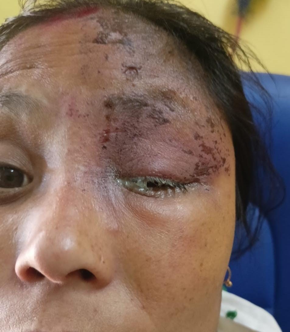

Figure 1: This image shows a left-sided HZO showing unilateral crusted lesions over the left forehead and periorbital region with associated upper-eyelid swelling
Image Courtesy: Captured by Author
Investigation
The diagnosis was primarily suspected clinically based on the characteristic unilateral vesicular and crusted eruption following the ophthalmic dermatome.
A comprehensive ocular assessment was performed to identify any associated ocular complications. Slit-lamp examination with fluorescein staining, intraocular pressure assessment, and dilated fundus examination were carried out.
No evidence of keratitis, anterior uveitis, or posterior segment involvement was detected at the time of examination.
Diagnosis and Clinical Reasoning
Based on the patient’s history and clinical examination, a diagnosis of left-sided HZO without significant ocular involvement at the time of presentation was made.
The unilateral distribution of the vesicular and crusted rash over the ophthalmic dermatome, together with the associated periocular symptoms, supported the clinical diagnosis.
Although no significant ocular involvement was identified initially, HZO can affect the eyelids, conjunctiva, cornea, and intraocular structures. Therefore, the absence of ocular complications at the initial examination does not eliminate the possibility of subsequent ocular involvement. Careful follow-up is consequently important. (1)
Management
The patient was treated with oral acyclovir 800 mg, five times daily for 7 days, along with oral analgesics for symptomatic relief.
Eye lubricants were prescribed to help prevent ocular dryness. The patient was advised not to rub or scratch the affected skin lesions.
Ophthalmic evaluation and follow-up were recommended because of the potential for delayed ocular complications associated with HZO.
At the one-week follow-up, the rash had progressed to crusted sores that were beginning to heal. No new ocular manifestations were identified, and visual acuity in the left eye remained 6/6.
Discussion
HZO occurs following reactivation of the varicella-zoster virus involving the ophthalmic division of the trigeminal nerve. The characteristic unilateral dermatomal eruption is an important clinical feature that facilitates diagnosis. (1,2)
Ocular involvement can range from relatively mild conjunctival inflammation to more significant complications such as keratitis and uveitis. In severe cases, deeper ocular structures may also become involved. (1,2)
Although the risk of ocular complications increases with age, HZO can also occur in younger adults, as demonstrated by this 35-year-old patient. (4,5)
In the present case, the patient had prominent cutaneous and periocular manifestations but no evidence of keratitis, anterior uveitis, or posterior segment involvement during the initial examination. This emphasises the importance of performing a complete ocular examination even when the patient’s primary complaint appears to be related to the skin.
Antiviral treatment is an important component of management, and continued ophthalmic follow-up is necessary because ocular inflammation may develop later or recur after the initial episode. (3)
The case therefore demonstrates the important role of Optometric and Ophthalmic assessment in patients presenting with periocular herpes zoster. Early recognition, appropriate treatment, patient education, and follow-up can help identify ocular complications at an early stage.
Conclusion
HZO can initially present predominantly with unilateral periocular skin manifestations while ocular structures remain unaffected. In this case, the characteristic left-sided ophthalmic dermatome involvement supported the diagnosis, while comprehensive ocular examination confirmed the absence of significant ocular complications at presentation.
The case highlights that a seemingly cutaneous presentation should not be considered solely a dermatological condition. Careful ocular examination and appropriate follow-up are essential because ocular complications may develop subsequently. Early antiviral therapy, symptomatic management, patient education, and continued ophthalmic monitoring remain important components of care.
References
- Litt J, Cunningham AL, Arnalich-Montiel F, Parikh R. Herpes zoster ophthalmicus: presentation, complications, treatment, and prevention. Infect Dis Ther. 2024;13(7):1439-59.
- Tuft S. How to manage herpes zoster ophthalmicus. Community Eye Health. 2020;33(108):71.
- Lo DM, Jeng BH, Gillespie C, Wu M, Cohen EJ. Current practice patterns and opinions in the management of recent onset or chronic herpes zoster ophthalmicus of Zoster Eye Disease Study investigators. Cornea. 2019;38(1):13-7.
- Yawn BP, Wollan PC, St Sauver JL, Butterfield LC. Herpes zoster eye complications: rates and trends. Mayo Clin Proc. 2013;88(6):562-70.
- Harding SP, Lipton JR, Wells JC. Natural history of herpes zoster ophthalmicus: predictors of postherpetic neuralgia and ocular involvement. Br J Ophthalmol. 1987;71(5):353-8.
About the Authors


Haziel Rynjah
Assistant Professor,
The Assam Royal Global University, Guwahati, India.

Jeba Ahmed
M. Optom Student,