


Syed Haseeb ur Rahman, B. Optom, PGDOVS
Consultant Optometrist, LV Prasad Eye Institute, Hyderabad, India
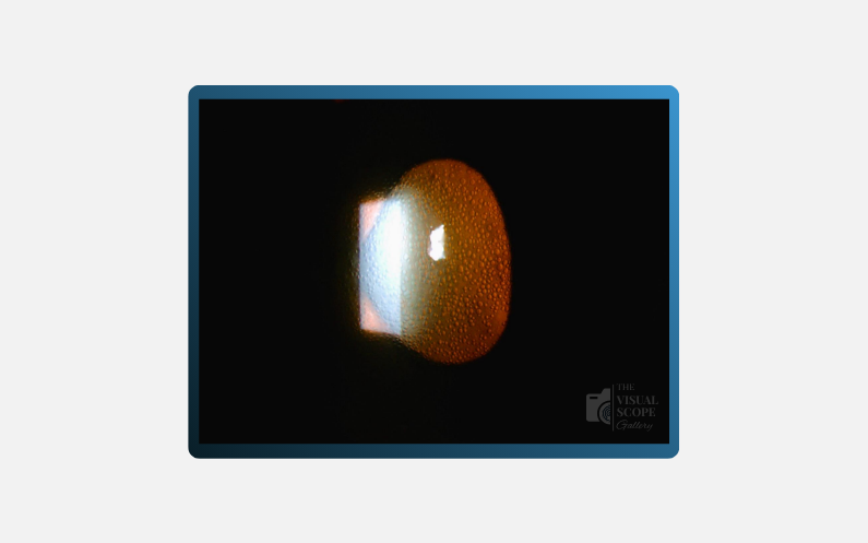

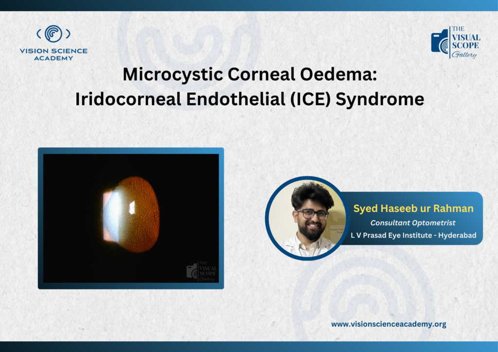
Figure: When the Corneal Endothelium Fails, Fluid Speaks: Microcystic Oedema in ICE Syndrome Captured in Light.
Image Summary
Slit-lamp photograph demonstrating diffuse microcystic epithelial corneal oedema in a patient with Iridocorneal Endothelial (ICE) syndrome. The characteristic epithelial microcysts result from endothelial dysfunction and failure of the corneal pump mechanism, leading to stromal and epithelial fluid accumulation. In ICE syndrome, abnormal endothelial cell proliferation and migration onto the trabecular meshwork and iris surface cause progressive Peripheral Anterior Synechiae (PAS), impairing aqueous outflow. This frequently results in secondary angle-closure glaucoma with elevated Intraocular Pressure (IOP), further exacerbating corneal oedema and visual disturbance. The presence of microcystic oedema may therefore serve as a clinical indicator of endothelial decompensation and uncontrolled IOP in ICE-related glaucomatous disease.