Kristi Sharma, M. Optom.
Senior Manager – Advanced Education and Partnerships, Vision Science Academy
Keywords
Retina; coloboma;contact lens; personalised treatment; rehabilitation
Declaration of Interest
This article is written purely for educational purpose. There are no conflicts of interest associated with the preparation or publication of this case report. No financial, commercial, or personal relationships influenced the management of the patient, interpretation of the findings, or preparation of the manuscript.
When a patient walks in to your clinic with an evident iris coloboma, what would your first thought be? Probably, you would think of a chorio-retinal coloboma that might be associated with it. Once you diagnose a chorio-retinal coloboma, what would your second thought be? Probably that colobomas are congenital conditions with no treatment. However, would you convey this generic information to the patient and say no treatment exists, or would you look deeper?
What is a Coloboma?
A coloboma is a congenital condition where a tissue of the eye does not form, or form partially during pregnancy. An iris coloboma is when a part of the iris goes missing or forms partially, and commonly occurs together with a chorio-retinal coloboma. The interesting detail is the fact that it is usually associated with early cataractous changes of the crystalline lens as well. (1)
In this case, we explore a typical story of iris and chorio-retinal coloboma. What was not typical in this case was how the patient’s chief complaint was managed with a simple personalised plan while the condition still remained incurable.
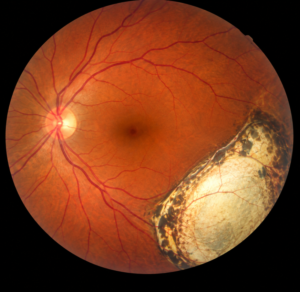

Figure 1: This image depicts the typical retinal appearance during Fundus Photography in a case of chorio-retinal coloboma.
Image Courtesy: Created by Author using Open AI
Case Overview
A 43-year-old male had been diagnosed with a premature cataract in the right eye at the age of 40 years and subsequently underwent cataract extraction with intraocular lens implantation. Following the procedure, the patient noticed that vision in the operated eye remained severely impaired. Even one week after surgery, he reported an inability to see adequately through the right eye.
Because of the persistent visual complaint, he presented to an eye-care clinic for further evaluation. Slit-lamp examination revealed an iris coloboma in the right eye. In view of this finding and the significant reduction in visual function, the patient was referred to a tertiary eye-care centre for detailed retinal evaluation.
At the tertiary centre, a comprehensive ocular and refractive assessment was performed. Visual acuity was recorded as counting fingers at 1 metre in the right eye and 6/6 in the left eye. The right eye was pseudophakic. Detailed fundus examination revealed a chorio-retinal coloboma involving the right eye. No other significant retinal abnormalities were identified on the initial examination.
The patient was counselled that coloboma represents a congenital ocular malformation rather than an acquired disease and that there was no specific treatment capable of reversing the underlying structural defect. However, the patient remained concerned about his inability to use the vision in his right eye and subsequently sought a second opinion at another tertiary eye-care centre.
During the subsequent evaluation, additional investigations, including Optical Coherence Tomography (OCT) and fundus photography, were performed. Importantly, these investigations demonstrated that the macular region was spared from the colobomatous defect. This finding suggested that despite the extensive structural anomaly, the eye retained potentially useful central visual function.
Based on the preserved macular anatomy, a contact lens trial was undertaken to determine whether optical rehabilitation could improve the patient’s functional vision. The trial resulted in a substantial improvement in visual acuity, from counting fingers at 1 metre to 6/24.
The improvement was clinically meaningful to the patient, who reported considerably better visual function through the affected eye. Following the successful trial, a prosthetic contact lens (C type) with a clear pupillary aperture was prescribed. The lens incorporated appropriate refractive correction within the pupillary zone while the prosthetic component helped address the excessive light entry and glare associated with the iris coloboma.
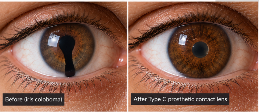

Figure 2: This image compares between an eye with coloboma and an eye with Type C prosthetic contact lens.
Image Courtesy: Created by Author using OpenAI
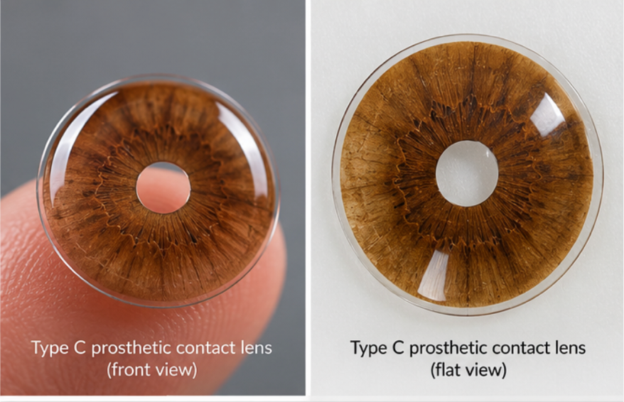

Figure 3: This image shows the general appearance of a Type C prosthetic contact lens.
Image Courtesy: Created by Author using OpenAI
The patient tolerated the contact lens well and reported significant satisfaction with the resulting visual improvement. He continues to use the prescribed contact lens as part of his visual rehabilitation.
Discussion
This case illustrates an important principle in clinical practice: the diagnosis and the patient’s complaint are not always the same problem.
In this patient, the diagnosis was ocular coloboma, a congenital condition for which there is no treatment that can restore the malformed ocular structures to normal. (1,2) However, the patient’s primary complaint was not the existence of the coloboma itself. His concern was functional: he was unable to see adequately through his right eye following cataract surgery.
The initial counselling that there was no treatment for coloboma was factually appropriate with respect to correcting the congenital structural anomaly. Nevertheless, from a patient-centred perspective, this does not necessarily represent the endpoint of management. The subsequent evaluation demonstrated that the macula was anatomically spared. This finding changed the clinical question from “Can the coloboma be treated?” to “How much useful visual function can this eye achieve, and how can that function be individua?”
This change in perspective was critical.
The contact lens trial served as a relatively simple but clinically informative functional assessment. The improvement from counting fingers at 1 metre to 6/24 demonstrated that the patient’s poor visual function was not solely attributable to irreversible retinal involvement. The preserved macular anatomy provided a basis for exploring optical rehabilitation.
The use of a prosthetic contact lens also addressed another important component of the patient’s symptoms. An iris defect can result in abnormal light entering the eye and may contribute to glare, photophobia, and reduced visual quality. A prosthetic lens with a controlled pupillary aperture can therefore serve both a cosmetic and functional purpose, while incorporation of refractive correction can further improve the available visual potential. (3)
The case consequently demonstrates the value of individualised management. Two patients with the same broad diagnosis of coloboma may have markedly different functional requirements depending on the anatomical structures involved, the extent of the defect, refractive status, symptoms, and personal visual demands.
It is therefore important that clinicians avoid equating “incurable” with “untreatable.” While the underlying congenital malformation may remain permanent, associated symptoms and functional limitations may still be addressed.
Conclusion
This case reinforces a fundamental principle of patient-centred eye care: we should treat the patient and their functional complaint, not merely the diagnosis.
Although ocular coloboma is a congenital structural anomaly for which there is no definitive curative treatment, comprehensive assessment may reveal preserved visual potential that can be improved through individualised rehabilitation. In this patient, recognition that the macula was spared led to a contact lens trial and subsequently to successful prosthetic contact lens management, with visual acuity improving from counting fingers at 1 metre to 6/24 and significant subjective satisfaction.
The case therefore highlights that the clinical responsibility does not necessarily end when we determine that a condition is incurable. Rather, it may begin with asking a different question: if we cannot change the condition, what can we still change for the patient?
For clinicians, this approach encourages a shift from disease-centred thinking toward functional, individualised and patient-centred care, where meaningful improvement may be achieved even when the underlying condition itself cannot be cured.
References
- Lingam, G., Sen, A. C., Lingam, V., Bhende, M., Padhi, T. R., & Xinyi, S. (2021). Ocular coloboma—a comprehensive review for the clinician. Eye, 35(8), 2086-2109.
- Singh, K., Snehi, S., Pattebahadur, R., & Kandi, M. (2024). Peering into the depths: chorio-retinal coloboma and risk of retinal detachment. BMJ Case Reports, 17(12), e262978.
- Srinivasan, F. S., Ting, M. D. S., Snyder, M. M. E., & Koch, M. H. R. (2014). Prosthetic iris devices. Canadian Journal of Ophthalmology, 49(1), 6-17.
About the Author