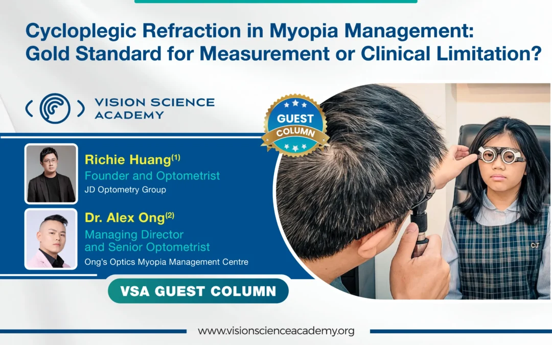
Richie Huang(2), (2)Founder and Optometrist, 2JD Optometry Group
Dr. Alex Ong(2), Managing Director and Optometrist, Ong’s Optics Myopia Management Centre
Cycloplegic refraction has long been regarded as the gold standard for paediatric refractive assessment because it minimises accommodative interference and reveals latent refractive components. However, its role in real-world Myopia Management remains debated, particularly in regions where Optometrists do not have access to cycloplegic agents. This paper critically evaluates whether cycloplegic refraction should be considered an indispensable requirement for clinical Myopia Management or primarily a tool for structural measurement and research standardisation. While cycloplegia improves refractive accuracy, it alters physiological visual conditions, including accommodation, pupil size, retinal image quality, and binocular interaction. These alterations limit its ability to represent habitual visual function. International clinical data further suggest that non-cycloplegic refraction, although less precise structurally, may better reflect functional vision and prescribing relevance in established Myopia. This paper proposed that cycloplegic refraction remains essential for baseline diagnosis, Hyperopia detection, Pseudo-Myopia differentiation, and research standardisation. However, in the ongoing management of established Myopia, it should not be regarded as an absolute requirement at every clinical encounter. A comprehensive model of Myopia Management should integrate structural refractive findings, subjective clarity, binocular vision status, axial length progression, and lifestyle-related risk factors.
Introduction
The global increase in Myopia prevalence has intensified the need for accurate refractive assessment and effective long-term management strategies in children. Cycloplegic refraction has traditionally been considered the gold standard in paediatric populations because it suppresses accommodation and improves the detection of latent Hyperopia and Pseudo-Myopia. (1,2)
However, real-world clinical practice is heterogeneous. In many countries, Optometrists do not have authorisation to administer cycloplegic agents, necessitating reliance on non-cycloplegic techniques. At the same time, modern Myopia Management extends beyond refractive measurement to include axial length monitoring, binocular vision status, accommodative function, visual behaviour, and environmental risk factors.
This raises a clinically important question:
Is cycloplegic refraction essential for every stage of Myopia Management, or should it be viewed primarily as a structural measurement tool within a broader clinical framework?
Cycloplegic vs Non-Cycloplegic Refraction: Structural vs Functional Information
Non-cycloplegic refraction in children is well known to overestimate Myopia due to residual accommodation, with reported differences of approximately 0.50–0.80 dioptres. (3)
As such, cycloplegic refraction remains the reference standard for determining refractive error in paediatric populations.
However, these two approaches represent different layers of clinical information:
- Cycloplegic refraction → structural refractive state
- Non-cycloplegic refraction → functional visual state
Importantly, subgroup analyses indicate that in established Myopic children, the difference between cycloplegic and non-cycloplegic measurements is often smaller (approximately 0.20–0.40 D), suggesting that the clinical impact on prescribing may be limited in this group. (4)
To facilitate clinical interpretation, key comparative findings from international studies are summarised in Table 1.
| Study | Population | Key Finding | Magnitude of Difference | Clinical Interpretation |
|---|---|---|---|---|
| Wilson et al., 2022 (3) | Children ≤12 yrs | Non-cycloplegic overestimates Myopia | ~0.50–0.80 D | Cycloplegia improves measurement accuracy |
| Sankaridurg et al., 2010 (5) | Myopic children | A smaller difference in established Myopia | ~0.20–0.40 D | Clinically less significant in Myopes |
| He et al., 2004 (6) | School children | Significant overestimation without cycloplegia | Up to >1.00 D | Critical for diagnosis |
| Rose et al., 2008 (7) | 6–12 yrs | The difference decreases with age | 0.84–1.18 D | Accommodation stronger in younger children |
Table 1: This table shows cycloplegic vs non-cycloplegic refraction (International Evidence).
From Measurement to Prescription: The Role of Functional Validation
Cycloplegic refraction provides a valuable baseline for structural refractive assessment. However, prescribing spectacles requires integration of subjective clarity, visual comfort, binocular coordination, and near visual demand.
Clinical guidelines recommend subjective refinement following cycloplegic examination, emphasising that cycloplegic findings should not be used in isolation. (1)
Studies on Post-Mydriatic Testing (PMT) demonstrate minimal differences in many Myopic children. (8,9) Furthermore, non-cycloplegic subjective refraction may better reflect habitual visual function and is therefore clinically relevant for prescribing. (9)
Physiological and Optical Limitations of Cycloplegia
Cycloplegia alters several key physiological and optical parameters:
- Elimination of accommodation
- Pupil dilation
- Reduced depth of focus
- Altered retinal image quality
These changes create a pharmacologically modified visual environment that differs from natural viewing conditions.
Clinical Strengths and Limitations of Cycloplegic Refraction
| Domain | Strengths | Limitations |
|---|---|---|
| Refractive Measurement | Eliminates accommodation, reveals latent error | Non-physiological state |
| Prescribing | Provides a structural baseline | Does not reflect functional clarity |
| Binocular Vision | Reduces accommodative variability | Alters AC/A and phoria |
| Optical Quality | Improves objectivity | Pupil dilation alters image quality |
| Clinical Use | Essential for diagnosis | Not always required for follow-up |
Table 2: This table shows cycloplegic refraction, the strengths vs limitations in clinical practice.
Binocular Vision: A Critical but Often Overlooked Dimension
Cycloplegia disrupts the accommodation–vergence relationship and may induce esophoric shifts and AC/A changes. (10) Thus:
- Binocular findings under cycloplegia do not represent real-world functional vision.
- This is especially important in Myopia Management, where near work demand and visual fatigue are key clinical concerns (11).
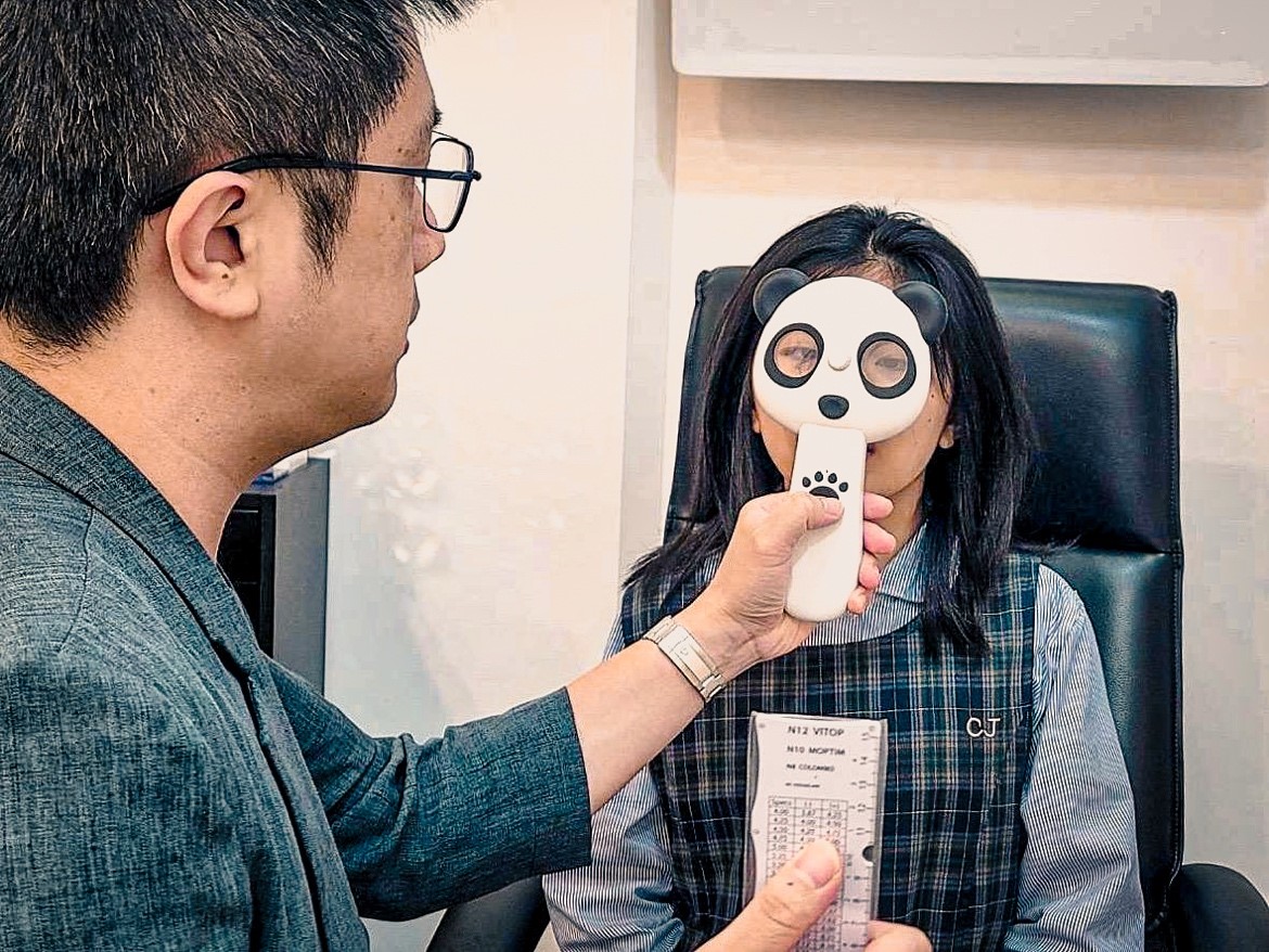
Picture 1: Binocular Accommodative Facility (BAF) Test using an automatic flipper of +/- 1.50D.
Image Courtesy: Capture by the Author
Under Correction: Functional Blur and Clinical Risk
Cycloplegic refraction often yields a less Myopic (less minus) result compared to non-cycloplegic measurements due to the elimination of accommodation. (4)
If prescribed without functional refinement, this may result in residual blur under natural viewing conditions once accommodation returns, as cycloplegic findings do not reflect habitual accommodative and binocular visual states. (1, 10) This raises an important clinical consideration:
Can prescribing directly from cycloplegic refraction lead to a form of functional under-correction?
Although cycloplegic refraction provides a structurally accurate baseline, prescribing must ultimately ensure clear retinal image quality under habitual conditions. (6) If the final correction leaves persistent distance blur, it may functionally resemble under-correction, which has been associated with accelerated Myopia progression. A randomised clinical trial demonstrated faster progression in under-corrected Myopic children compared to those receiving full correction. (12) Consistent with this, the International Myopia Institute does not recommend under-correction as a Myopia control strategy, emphasising the importance of providing full and clear optical correction to minimise retinal defocus. (2)
It is important to distinguish that cycloplegic prescribing is not inherently equivalent to intentional under-correction. In many established Myopic children, the difference between cycloplegic and non-cycloplegic refraction is relatively small (approximately 0.20–0.40 D) and may remain clinically acceptable.(13) However, when residual blur is present, the potential for retinal defocus under real-world conditions must be considered.
Therefore, cycloplegic refraction should be interpreted as a structural reference rather than a prescribing endpoint. Functional validation through subjective refinement and binocular vision assessment is essential to ensure that the final prescription achieves full, clear, and sustainable vision, minimising the risk of progression associated with persistent defocus.
Reframing the “Gold Standard”
Cycloplegic refraction is widely regarded as the gold standard for the measurement of refractive error in children, as it minimises accommodative interference and provides a closer approximation of the structural refractive state of the eyes. (1,2,4)
A gold standard for measurement does not necessarily translate into a gold standard for prescribing or comprehensive Myopia Management. Spectacle prescribing requires consideration of functional visual performance under habitual conditions, including subjective clarity, accommodative response, binocular coordination, and visual comfort. Cycloplegia, by altering accommodation, pupil size, and the accommodation–vergence relationship, does not fully represent these real-world visual conditions. (11)
Accordingly, clinical guidelines recommend that cycloplegic findings be interpreted alongside subjective refinement rather than used in isolation. (1)
Importantly, the absence of cycloplegic refraction does not imply that refractive assessment is inaccurate or unsuitable for Myopia Management. Well-conducted non-cycloplegic refraction, when combined with subjective refinement and binocular vision evaluation, can provide clinically meaningful and functionally relevant information for prescribing, particularly in children with established Myopia. Contemporary Myopia Management extends beyond refractive error alone, incorporating axial length monitoring, visual behaviour, and environmental risk factors. (2) Therefore, while cycloplegic refraction remains an essential reference for structural assessment, it should be regarded as one component within a broader, integrated clinical framework rather than the sole determinant of prescribing or management decisions.
Global Clinical Reality and Practical Model
In real-world clinical practice, access to cycloplegic refraction varies widely due to differences in regulatory scope, resource availability, and clinical workflow. Despite this variability, effective Myopia Management continues to be achieved globally through a combination of subjective refraction, binocular vision assessment, axial length monitoring, and lifestyle modification. These approaches reflect the multifactorial nature of Myopia progression, which is influenced not only by refractive error but also by accommodative behaviour, visual demand, and environmental exposure. (2)
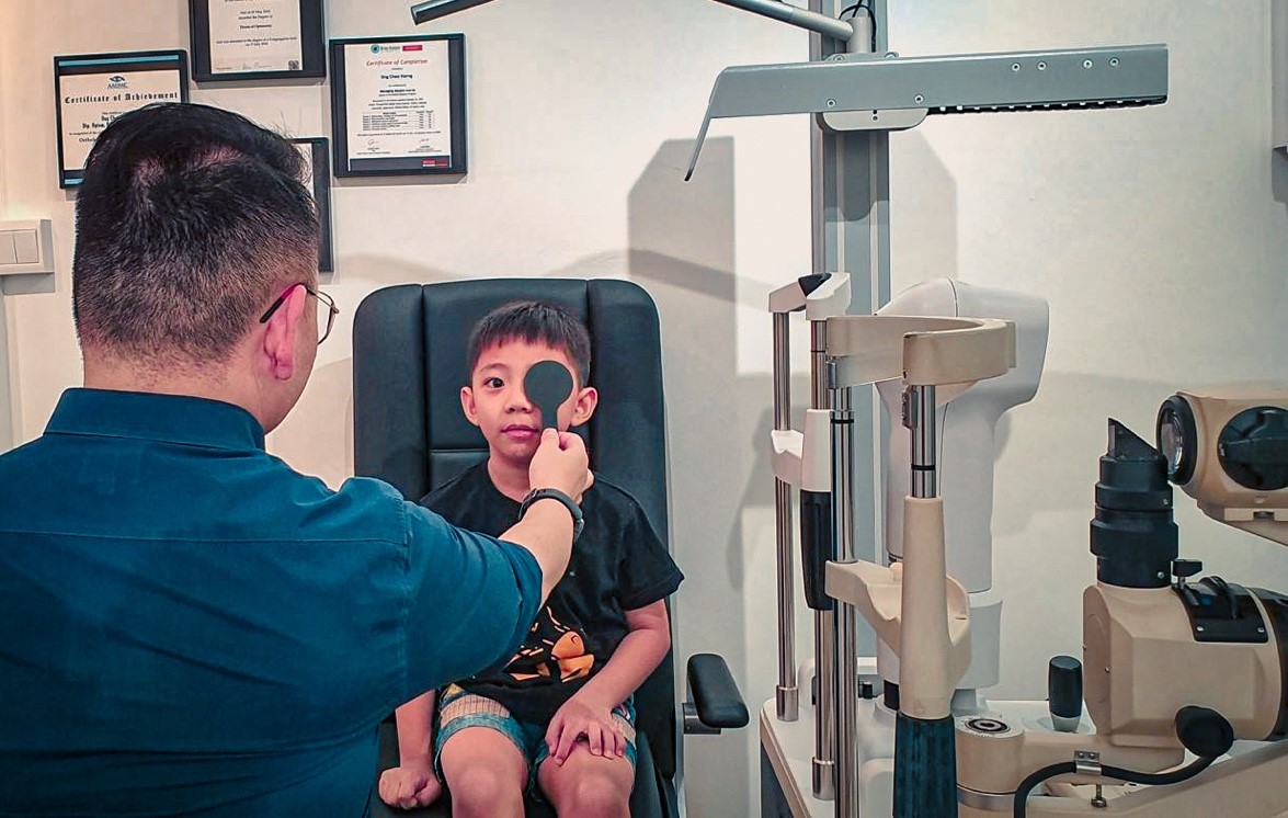
Picture 2: Distance cover test check for phoria or tropia
Image Courtesy: Created by the Author
Contemporary clinical frameworks, including those proposed by the International Myopia Institute, emphasise longitudinal monitoring and risk factor modification alongside optical correction, rather than reliance on a single measurement method. In patients with established Myopia who are cooperative and regularly followed, non-cycloplegic refraction combined with functional and binocular vision assessment can provide clinically meaningful guidance for prescribing and management. Therefore, a practical model of Myopia care should prioritise the integration of structural baseline measurements, functional visual performance, binocular coordination, and progression tracking, as summarised in Table 3, rather than depending solely on the availability of cycloplegic refraction.
| Component | Measurement Method | Clinical Role |
|---|---|---|
| Structural Refraction | Cycloplegic | Baseline |
| Functional Refraction | Subjective | Prescribing |
| Binocular Vision | BV testing | Comfort |
| Progression | Axial length | Monitoring |
| Environment | Lifestyle | Risk control |
Table 3: This table highlights clinical decision-making model in Myopia Management.
Conclusion
Cycloplegic refraction remains an essential tool for paediatric refractive assessment and research standardisation, providing a reliable estimate of the structural refractive state by minimising accommodative interference. However, its role in clinical practice should be carefully contextualised. Because cycloplegia alters accommodation, pupil size, optical quality, and binocular interaction, it does not fully represent functional vision under habitual viewing conditions.
A clinically meaningful approach to Myopia Management requires integration of structural measurement with functional visual performance, binocular vision status, and longitudinal progression data. While cycloplegia provides an important foundation particularly in initial diagnosis and selected clinical scenarios it should not be overextended as a universal requirement for prescribing or ongoing management. Ultimately, effective Myopia care depends on a comprehensive, patient-centred framework rather than reliance on a single measurement standard.
References
- Hutchinson AK, Morse CL, Hercinovic A, Cruz OA, Sprunger DT, Repka MX, Lambert SR, Wallace DK; American Academy of Ophthalmology Preferred Practice Pattern Pediatric Ophthalmology/Strabismus Panel. Pediatric eye evaluations preferred practice pattern®. Ophthalmology. 2023;130(3): P222–P270.
- Gifford KL, Richdale K, Kang P, Aller TA, Lam CSY, Liu YM, Michaud L, Mulder J, Orr JB, Rose KA, Saunders KJ, Seidel D, Tideman JWL, Sankaridurg P. IMI – Clinical management guidelines report. Invest Ophthalmol Vis Sci. 2019;60(3):M184–M203.
- Wilson S, Ctori I, Shah R, Suttle C, Conway ML. Systematic review and meta-analysis on the agreement of non-cycloplegic and cycloplegic refraction in children. Ophthalmic Physiol Opt. 2022;42(6):1276–1288.
- Sankaridurg P, He X, Naduvilath T, Lv M, Ho A, Smith EL 3rd, Erickson P, Zhu J, Zou H, Xu X. Comparison of noncycloplegic and cycloplegic autorefraction in categorizing refractive error data in children. Acta Ophthalmol. 2017;95(7): e633–e640.
- Sankaridurg P, Donovan L, Varnas S, Ho A, Chen X, Martinez A, Fisher S, Lin Z, Smith EL 3rd, Ge J, Holden B. Spectacle lenses designed to reduce progression of myopia: 12-month results. Optom Vis Sci. 2010;87(9):631–641.
- He M, Zeng J, Liu Y, Xu J, Pokharel GP, Ellwein LB. Refractive error and visual impairment in urban children in southern China. Invest Ophthalmol Vis Sci. 2004;45(3):793–799.
- Rose KA, Morgan IG, Ip J, Kifley A, Huynh S, Smith W, Mitchell P. Outdoor activity reduces the prevalence of myopia in children. Ophthalmology. 2008;115(8):1279–1285. doi:10.1016/j.ophtha.2007.12.019.
- Khan T, Jahangir S. Authenticity of glasses prescribed after cycloplegic refraction as verified by post-mydriatic test. Ophthalmol Pak. 2018;8(2):24–28.
- Kothari M, Hussain A. Is post-mydriatic test necessary in children having compound myopic astigmatism? J Clin Ophthalmol Res. 2015;3(2):77–79.
- Asharlous A, Yekta A, Hashemi H, et al. The effect of cycloplegia on ocular alignment and accommodative convergence/accommodation ratio. BMC Ophthalmol. 2024;24:Article 89.
- Gwiazda J, Hyman L, Hussein M, Everett D, Norton TT, Kurtz D, Leske MC, Manny R, Marsh-Tootle W, Scheiman M. A randomized clinical trial of progressive addition lenses versus single vision lenses on the progression of myopia in children. Invest Ophthalmol Vis Sci. 2003;44(4):1492–1500.
- Chung K, Mohidin N, O’Leary DJ. Undercorrection of myopia enhances rather than inhibits myopia progression. Vision Res. 2002;42(22):2555–2559.
- Benjamin WJ. Borish’s clinical refraction. 2nd ed. St. Louis (MO): Butterworth-Heinemann; 2006.
About the Author

Mr. Richie Huang, Optometrist, FOWNS, CPNP (ANA)
B.Sc Optom Taiwan
Mr. Huang is a practicing Optometrist and Founder of JD Optometry Group in Taiwan. He is also the current President of the New Taipei City Optometrists Association and was the Past President of the Taiwan Optometrists Association (TOA). He specialises in elderly vision care and children’s vision development, combining clinical expertise with a strong commitment to professional education. His work bridges Visual Nutrition, Paediatric Myopia Management, Binocular Vision and Geriatric Vision Care, with strong engagement in public education and professional training. As an Optometrist and a Fellows of Ocular Wellness Nutrition Society, his passion for Optometry and Nutrition is reflected in his ongoing contributions to public awareness and his leadership in advancing eye care standards across Taiwan.

Dr. Alex Ong, DOptom, FAAO, FOWNS, CPNP (US)Optometrist, Singapore
PGCert (Anterior Eye Diseases, Cataract and Glaucoma)
PGCert (Retinal Disease Recognition and Co-Management)
MSCO, B.Sc Optom, Dip. Optom, Singapore
Dr. Ong is the Founder and Managing Director/Optometrist at Ong’s Optics Myopia Management Centre, established in 2005 and 2nd generation owner of Ong’s Optics and Contact Lens Centre, established in 1985. With over two decades of clinical experience, his expertise spans evidence-based Myopia Management, Ocular Nutrition, Binocular Vision, Orthokeratology, and Geriatric Vision Care. He serves as a Key Opinion Leader for Menicon Japan and has taught as an adjunct lecturer at both optometry schools in Singapore. Dr. Ong is actively involved in Optometric education and professional training across Asia. As a Certified Personalised Nutrition Practitioner (CPNP) with the American Nutrition Association, he integrates nutritional science with vision care to promote a proactive and preventive model of lifelong eye health. He is also a member of Ocular Wellness and Nutrition Society.
Recent Comments