


Dr. Sanjay Kumar Mishra1, Scientist
1Dr. R. P. Centre for Ophthalmic Sciences, All India Institute of Medical Sciences (AIIMS), New Delhi, India
Samyak Mishra2, B. Optom Student
2School of Allied Health Sciences and Research, Jamia Hamdard, New Delhi, India
Keywords
visual acuity; visual function; retinal disease; microperimetry; multifocal electroretinography
Declaration of Interest
The blog is written solely for education purpose, and it does not have any financial support and conflict of interest.
Seeing Beyond the Chart
Consider a patient with Diabetic Macular Oedema who reads the 6/6 line, yet, reports difficulty recognising faces in dim light, reading continuously, or seeing subtle detail. What, then, does “6/6” really tell us?
Visual acuity is essential, but it is only one measure of vision. A broader assessment pathway is illustrated in Figure 1. It primarily tests the ability to resolve small, high-contrast detail under standardised conditions. It does not capture the full range of visual demands encountered in daily life. (¹)
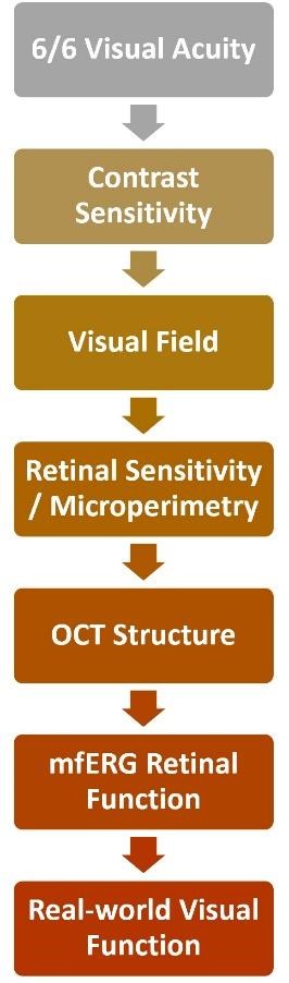

Figure 1: This flowchart shows the multidimensional assessment of visual function beyond visual acuity.
Vision Beyond the Chart
Everyday visual performance also depends on contrast sensitivity, luminance, visual field, fixation, and reading ability. Contrast sensitivity may reveal deficits that remain inconspicuous when high-contrast acuity is preserved. Patient-reported outcomes and task-based measures can add another layer of information about how retinal disease affects daily life. (¹–³)
This matters particularly in retinal disease. Two people with similar visual acuity may have different retinal sensitivity, fixation behaviour and functional difficulties.
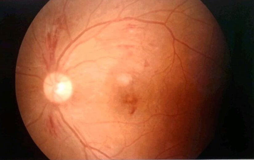

Figure 2: This is a representative colour fundus photograph of a patient with diabetic retinal disease.
Image Courtesy: Captured by Authors
The more useful question is therefore not simply, “How many letters can the patient read?” but, “How well is the visual system functioning across the retina?”
Looking Beyond the Retina’s Appearance
Optical Coherence Tomography (OCT) has transformed retinal care by allowing clinicians to visualise retinal structure in vivo. An example is shown in Figure 2.
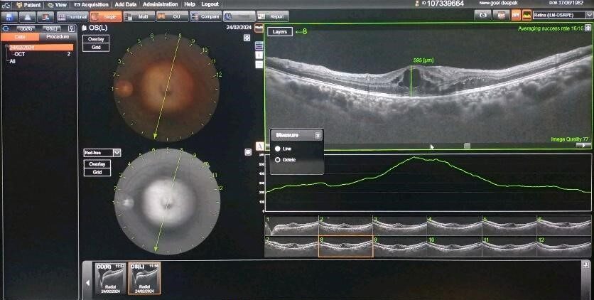

Figure 3: This is a representative macular OCT demonstrating retinal structural changes and central macular thickness.
Image Courtesy: Captured by Authors
Yet, anatomical improvement does not necessarily mean that every aspect of visual function has recovered. (1,4)
Microperimetry complements OCT by mapping retinal sensitivity at specific retinal locations while monitoring fixation. Figure 3 shows the clinical assessment.
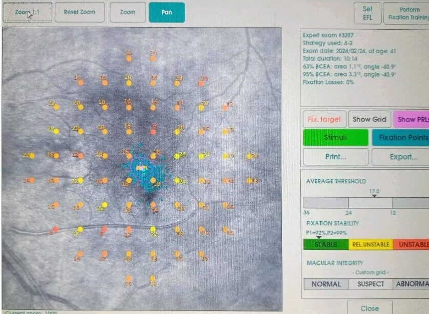

Figure 4: This is a representative microperimetry map showing spatial retinal sensitivity and fixation behaviour.
Image Courtesy: Captured by Authors
It can detect local functional impairment and has become increasingly relevant in the evaluation of macular diseases, including Diabetic Macular Oedema. (4,5) Recent evidence suggests that microperimetry can detect functional improvement after anti-vascular endothelial growth factor treatment even when best-corrected visual acuity shows little change. (6)
Multifocal electroretinography (mfERG) provides a different perspective by measuring localised electrical responses from the retina. Figure 4 illustrates the corresponding electrophysiological assessment.
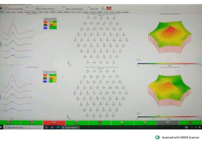

Figure 5: This is a representative mfERG recording demonstrating localised retinal electrophysiological responses.
Image Courtesy: Captured by Authors
In Diabetic Macular Oedema, mfERG abnormalities have been associated with visual acuity and OCT features, supporting the value of combining structural and functional assessment. (7)
From 6/6 to Meaningful Vision
The field is moving towards a multidimensional definition of visual function. Recent work in centre-involving Diabetic Macular Oedema has shown that retinal sensitivity may be more strongly associated with several vision-related quality-of-life domains than best-corrected visual acuity alone. (8) Newer studies also support microperimetry as a sensitive functional endpoint for detecting treatment-related change. (6,9)
This does not make visual acuity obsolete. It puts the acuity chart in its proper place: one measure within a broader assessment of retinal structure, function and patient experience.
For clinicians, the goal is not to replace 6/6, but to understand what lies behind it.
Because in retinal disease, the question may no longer be, “Can the patient see 6/6?”
It may be:
“Can the patient see, detect, locate and use visual information effectively in everyday life?”
References
- Ríos HA, Lövestam-Adrian M, Plainis S, Tsilimbaris M, Joussen AM, Keegan D, et al. Additional measures of macular function beyond visual acuity. Graefes Arch Clin Exp Ophthalmol. 2024;262(6):1723-1736. doi:10.1007/s00417-023-06272-1.
- Richman J, Spaeth GL, Wirostko B. Contrast sensitivity basics and a critique of currently available tests. J Cataract Refract Surg. 2013;39(7):1100-1106. doi:10.1016/j.jcrs.2013.05.001.
- Puell MC, Contreras I, Pinilla I, Escobar JJ, Soler-García A, Blasco AJ, et al. Beyond visual acuity: Patient-relevant assessment measures of visual function in retinal diseases. Eur J Ophthalmol. 2021;31(6):3149-3156. doi:10.1177/1120672121990624.
- Horie S, Corradetti G, Esmaeilkhanian H, Sadda SV, Cheung CMG, Ham Y, et al. Microperimetry in retinal diseases. Asia Pac J Ophthalmol (Phila). 2023;12(2):211-227. doi:10.1097/APO.0000000000000597.
- Rai BB, Maddess T, Nolan CJ. Functional diabetic retinopathy: A new concept to improve management of diabetic retinal diseases. Surv Ophthalmol. 2025;70(2):232-240. doi:10.1016/j.survophthal.2024.11.010.
- Deng WL, Liu KY, Li ZY, Zhang XD, Zhang YL, Liu SL. Baseline microperimetry and metabolic status predict functional outcomes in diabetic macular oedema: a prospective cohort study of anti-VEGF therapy. Ann Med. 2026;58(1):2687175. doi:10.1080/07853890.2026.2687175.
- Khojasteh H, Riazi-Esfahani H, Khalili Pour E, Faghihi H, Ghassemi F, Bazvand F, et al. Multifocal electroretinogram in diabetic macular edema and its correlation with different optical coherence tomography features. Int Ophthalmol. 2020;40:571-581. doi:10.1007/s10792-019-01215-4.
- Piccoli G, Fenwick EK, Bucceri V, Lamoureux EL, Tagliabue E, Nucci P, et al. Impact of vision impairment and microperimetry parameters on quality of life in patients with center-involving diabetic macular edema. Ophthalmol Sci. 2026;6(2):101001. doi:10.1016/j.xops.2025.101001.
- Menna F, Calabro M, Guillot A, De Luca L, Meduri A, Baldascino A, et al. Functional real-world outcomes of aflibercept 8 mg versus faricimab after loading phase in diabetic macular edema. Ophthalmol Ther. 2026;15(7):2245-2254. doi:10.1007/s40123-026-01395-0.
About the Authors


Dr. Sanjay Kumar Mishra
Scientist,
Dr. R. P. Centre for Ophthalmic Sciences, All India Institute of Medical Sciences (AIIMS), New Delhi, India

Samyak Mishra
B. Optom Student,