


Prof. Dr. Johannes (Hans) van der Steen
Chair of Visual Information Processing, Department of Neuroscience, Erasmus MC Rotterdam, The Netherlands
When I was a child at age 7, my parents gave me a Viewmaster: a plastic device with two lenses. On the top there was a slit where I could insert a cardboard rotating disc with slide images. The miracle happened when I held the device in front of both eyes and looked through it. Instantaneously beautiful scenes in full depth appeared. At the age of 75 the concept of 3D vision still intrigues me.
Looking at the world with two eyes seems effortless. We normally perceive a single, stable and three-dimensional visual environment, despite the fact that each eye receives an image from a slightly different viewpoint. The brain combines these two images into a unified percept using differences in retinal location on each retina (binocular disparity) to derive information about depth (stereopsis). In order to maintain binocular fixation, the eyes have to continuously perform highly coordinated movements.
From BC to the 19th Century
Questions about binocular vision are centuries old. Aristotle (ca 384-322 BC)
already wondered how the images of the two eyes result in a single visual percept.
Up to the 19th century the overriding concern was how the world is seen single with two eyes. Porta (1535–1615) advanced a theory of binocular single vision that maintained that we only see with one eye at one time. Two decades later, Franciscus Aguilonius (1567–1617; 1613) suggested an alternative interpretation, the images in the eyes are fused or combined. (see for a review on history binocular vision, Wade, 2021)
An important step was made when in 1838, Charles Wheatstone introduced the stereoscope, an instrument that presented slightly different images separately to the two eyes. When these images were appropriately combined by the brain, observers experienced a compelling perception of depth. This demonstrated that depth perception could arise from the small positional differences between the images projected onto the two retinas, known as binocular disparity. The stereoscope therefore became a powerful experimental tool for investigating stereoscopic vision.
Since that time the scientific study of the interaction between binocular eye movements, sensory fusion and stereopsis has evolved. From the nineteenth-century stereoscope to modern computational models of vergence, researchers have progressively uncovered how the visual system aligns the eyes and neuronal codes differences between retinal images into a perception of three-dimensional space.
The nineteenth century saw important theoretical contributions from scientists such as Hermann von Helmholtz and Ewald Hering. Concepts such as corresponding retinal points, the horopter and the hypothetical cyclopean eye, that were framed earlier became central to the theories of binocular vision. (see for a review on the origin of terms in binocular vision, Wade, 2021)
Although these ideas remained influential well into the twentieth century, they do not answer the key question of how we perceive a 3D image. More recent experimental work demonstrates that binocular perception is more complex than a simple geometric combination of the positions of the two eyes and their retinal images. For a more complete understanding of 3D-vision we must consider the role of binocular eye movements and the neural basis of depth vision.
Binocular Eye Movements: The Motor Foundation of Stereopsis
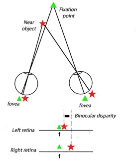
Stereopsis cannot be understood independently of ocular motor control. When we fixate an object, both eyes must rotate so that the object’s image falls approximately on corresponding retinal locations, ideally near the fovea. When the fixation distance changes, the eyes rotate in opposite directions (vergence). For a nearby object, the eyes rotate inward, producing convergence. For a distant object, less convergence is required. Vergence therefore provides an essential motor component of binocular vision.
An important study on ocular vergence was done in 1989 while observers looked at real three-dimensional targets at different distances and directions (Erkelens et al., 1989). Their studies demonstrated how vergence changes continuously when target distance changes and how vergence is combined with other components of eye movement during gaze shifts. This work was important because everyday gaze shifts rarely involve a change in only one variable. When we look from one object to another, the target may differ simultaneously in direction and distance. The visual system must therefore coordinate version, which moves both eyes in the same direction, with vergence, which moves the eyes in opposite directions.

The Neural Basis of Depth
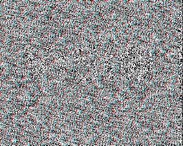
The introduction of random-dot stereograms by Bela Julesz (1971) led to the breakthrough that the visual system can extract stereoscopic depth from binocular disparity alone.
In these stimuli, recognisable contours and objects can be eliminated, leaving only small differences between two random patterns. When the two patterns are presented separately to the two eyes, a three-dimensional percept emerges.
Random dot stereograms have been used extensively to study the neural basis of depth vision. A common feature from animal studies is that binocular disparity selectivity in visual cortex V1 appears to shape the reliance on stereoscopic cues to interpret the world. Higher visual areas in primate are characterised by specialisation for 3D processing linked to stable perception and interacting with objects. In addition to stereopsis, perspective and other cues play prominent roles in 3D vision (Parker et al., 2022).
Recent non-invasive MRI studies in humans indicate a similar organisation, although it remains challenging to perform such studies (for a review see Rosenberg et al, 2025)
Depth Perception and Eye Movements
A particularly influential study published in 1985 examined eye movements and stereopsis during dichoptic viewing of moving random-dot stereograms (Erkelens and Collewijn, 1985a).. Their findings contributed to the understanding that vergence, disparity and the perception of motion in depth are related but distinct processes. Their experiments also showed that binocular fusion has limits. When the disparity between the two retinal images becomes sufficiently large, fusion can be lost, and the eyes can no longer maintain normal binocular correspondence. When the stimulus is subsequently brought back within the fusion range, binocular coordination can be regained (Erkelens and Collewijn, 1985b)
This distinction is scientifically important. A person may experience stereoscopic depth because of retinal disparity, while the eyes simultaneously respond with vergence movements. Yet neither the magnitude of vergence nor retinal disparity alone completely explains the resulting percept. Stereopsis is therefore better understood as the outcome of an interaction between sensory and motor mechanisms.

Clinical Significance: Strabismus and Amblyopia
The scientific understanding of binocular eye movements and stereopsis has direct clinical relevance. In strabismus, the visual axes of the two eyes are misaligned. This can interfere with binocular fusion and stereopsis. Particularly during childhood, the brain may suppress information from one eye to avoid double vision. Persistent abnormal binocular experience can contribute to amblyopia.
Stereopsis is consequently an important indicator of binocular visual function. Clinical stereotests measure the smallest binocular disparity that a person can detect as a difference in depth. However, stereopsis should not be interpreted in isolation. Ocular alignment, visual acuity, ocular motility, sensory fusion and functional visual performance all contribute to a complete assessment. Binocular vision is not merely a matter of whether the eyes appear aligned. The dynamic coordination of the two eyes and the way visual information is processed by the brain are equally important.
Why Stereopsis Remains Important
Stereopsis contributes to the perception of relative depth and spatial relationships. It is particularly valuable when fine depth discrimination is required. Nevertheless, human depth perception does not depend exclusively on stereopsis. Perspective, shading, motion, occlusion and other monocular cues also provide information about three-dimensional structure.
This is one reason why the relationship between binocular and monocular vision has become an increasingly important topic in modern vision science. The history of binocular vision is therefore a progression from optical demonstrations to increasingly sophisticated models of perception and neural control. Wheatstone demonstrated that binocular disparity can create a compelling sense of depth. Helmholtz and Hering developed influential theories of binocular vision. Julesz demonstrated that stereopsis can emerge from random-dot patterns. Later neurophysiological research identified neural mechanisms sensitive to binocular disparity. Erkelens et al. demonstrated the interaction between eye movements and depth perception.
The broader lesson from almost two centuries of research is that seeing with two eyes is not simply a matter of adding two retinal images together. It is a continuous dialogue between eye movements, retinal disparity, neural processing, visual direction and the three-dimensional environment.
References
- Wade NJ. On the Origins of Terms in Binocular Vision. Iperception. 2021 Feb 24;12(1):2041669521992381. doi: 10.1177/2041669521992381. PMID: 33717428; PMCID: PMC7926055.
- Wheatstone, C. (1838). Contributions to the physiology of vision—Part the first. On some remarkable, and hitherto unobserved, phenomena of binocular vision. Philosophical Transactions of the Royal Society, 128, 371–394.
- Julesz B. Foundations of Cyclopean Perception. Chicago University Press; Chicago, IL, USA: 1971.
- Erkelens CJ, Collewijn H. Eye movements and stereopsis during dichoptic viewing of moving random-dot stereograms. Vision Res. 1985;25(11):1689-700. doi: 10.1016/0042-6989(85)90141-5. PMID: 3832593.
- Erkelens CJ, Collewijn H. Eye movements in relation to loss and regaining of fusion of disjunctively moving random-dot stereograms. Hum Neurobiol. 1985;4(3):181-8. PMID: 4066427.
- Ari Rosenberg, Holly Bridge, Charles E. Connor, Jason M. Samonds, Seiji Tanabe, Kristine Krug. The Richness of Vision in the Mammalian Brain: Neural Codes for Visual Perception and Behavior in Three Dimensions. Journal of Neuroscience 12 November 2025, 45 (46) e1316252025; DOI:10.1523/JNEUROSCI.1316-25.2025
About the Author


Hans van der Steen was until 2021 Full Professor at the department of Neuroscience, Erasmus MC Rotterdam, The Netherlands. He held the chair on visual information processing at Erasmus University endowed by Royal Dutch Visio (a national Vision rehabilitation center).
After his biology study at Utrecht University he did his PhD training in Rotterdam under the supervision of Prof. Han Collewijn at Erasmus University Rotterdam and continued his international training at various prestigious institutions such as University College, London, UK (Royal Society Fellowship) and the National Eye Institute (NIH) in Bethesda, USA. (human frontiers program). He has supervised a team of 12 researchers, PhD students and Research Master students. He has (co-) published in total 120 research papers and book chapters.
In his research technical advances in the precise monitoring of eye movements have become increasingly important in the diagnosis and monitoring of visual sensory loss by neurological damage or disease. In close collaboration with clinical departments and national visual rehabilitation centers, his team has used eye tracking technology to study vestibular problems in adults and children. His work has led to several innovative technologies to detect cerebral visual pathologies at an early stage of the disease. In this endeavor his team still works together with national teams, in particular Royal Dutch Visio, Neonatology, Neurology, ENT, Ophthalmology and Geriatrics at Erasmus MC, and with Technical University Delft. International collaborations have been set-up with Sankara Nethralaya, Chennai, India, Belgium, Germany, Uk, Sweden and Norway.