


Ritika Jain, B. Optom.
M. Optom Student, CT University, Ludhiana, India
The tight relationship between the brain and eyes is demonstrated by the fact that the retina and optic nerve are extensions of brain tissue. Therefore, neurological conditions like Alzheimer’s Disease (AD) and Dementia can also affect the eyes. Researchers are looking into this connection to better understand and perhaps detect brain diseases through eye exams. (1)
Dementia is defined as a significant reduction in cognitive function that is severe enough to interfere with daily functioning. Of the several types of Dementia, AD is the most common, accounting for over two-thirds of cases in persons 65 and older. Alzheimer’s is a progressive neurological illness that impairs judgment, reasoning, language, memory, comprehension, and concentration. (2)
Can the Eyes Reveal Alzheimer’s?
Alzheimer’s Disease, a chronic, progressive neuro-degenerative disease, is increasingly associated with retinal biomarkers because the retina remains in embryological and anatomical continuity with the Central Nervous System (CNS). The retina develops typical AD pathologies such as tau deposits and amyloid-beta plaques, with microvascular alterations and thinning of the nerves. These structural and vascular changes are detected by imaging instruments such as Optical Coherence Tomography (OCT) and OCT Angiography (OCTA). Amyloid detection is also improved by newer technologies, including hyperspectral imaging and confocal scanning laser ophthalmoscopy. A combination of these ophthalmological examinations enables pre-clinical AD diagnosis, enabling earlier treatment and better patient management. (3)
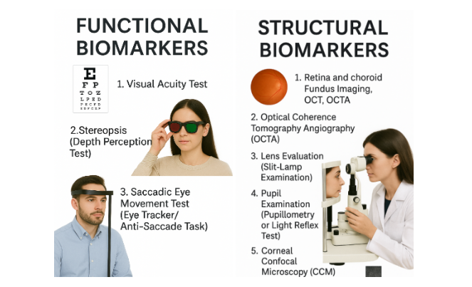

Figure 1: Structural vs Functional Biomarkers
Structural Biomarkers
There are anatomical or physiological changes in ocular structures that may indicate AD. (4)
1. Retina and Choroid
Examinations: Retinal fundus photography, OCT, and OCTA.
a. OCT (Optical Coherence Tomography) Findings: Retinal inclusion bodies, Ganglion Cell – Inner Plexiform Layer (GC-IPL) decrease, and Retinal Nerve Fibre Layer (RNFL) thinning. Macular and peripapillary regions are frequently studied.
b. OCTA (OCT Angiography) Findings: Reduced superficial and deep vascular density (VD), particularly in the parafoveal and macular regions, and an increased Foveal Avascular Zone (FAZ) area are all indicative of early AD-related damage. (4)
2. Lens Evaluation (Slit-Lamp Examination)
a. Findings: Mis-folded protein aggregates in AD; however, amyloid-beta presence is low or absent, making it a less specific marker. Also, observe for the presence of cataracts.
3. Pupil Examination (Pupillometry or Light Reflex Test)
a. Findings: Larger pupil size, abnormal light reflex, altered response to cholinergic agents due to acetylcholine deficiency, a hallmark of AD.
4. Corneal Confocal Microscopy (CCM)
a. Findings: Decreased length, density, and branching of nerve fibres. Strong association with cognitive impairment. (4)
Functional Biomarkers
These tests assess visual and cognitive processing performance. (4)
- Visual Acuity: May remain normal; declines in low light may indicate early dysfunction.
- Stereopsis: Depth perception deficits reflect impaired visuo-spatial processing.
- Saccadic Eye Movements: Delayed and error-prone anti-saccades suggest early frontal lobe involvement. (4)
Conclusion
Imaging technology, such as OCT, OCTA, and other sophisticated imaging machines, now enables eye health specialists to detect subtle warning signs, such as nerve thinning or decreased blood flow, that may be a sign of Alzheimer’s, even before there are symptoms of memory loss. By monitoring these changes with routine eye exams, optometrists can be a great help to patients by providing them early treatment and support.
References
- Kate Rauch. (2022). Alzheimer’s Disease, Dementia and the Eye. [online] American Academy of Ophthalmology. Available at: https://www.aao.org/eye-health/diseases/alzheimers-disease-dementia-eye#:~:text=Alzheimer’s%20Disease%2C%20Dementia%20and%20the,Alzheimer’s%20Disease%20Through%20the%20Eye?
- Kumar, A., Sidhu, J., Lui, F. and Tsao, J.W. (2024). Alzheimer Disease. [online] Nih.gov. Available at: https://www.ncbi.nlm.nih.gov/books/NBK499922/#:~:text=Pathophysiology,basal%20forebrain%20and%20the%20neocortex.
- Gaire, B. P., Koronyo, Y., Fuchs, D. T., Shi, H., Rentsendorj, A., Danziger, R., Vit, J. P., Mirzaei, N., Doustar, J., Sheyn, J., Hampel, H., Vergallo, A., Davis, M. R., Jallow, O., Baldacci, F., Verdooner, S. R., Barron, E., Mirzaei, M., Gupta, V. K., Graham, S. L., … Koronyo-Hamaoui, M. (2024). Alzheimer’s disease pathophysiology in the Retina. Progress in retinal and eye research, 101, 101273. https://doi.org/10.1016/j.preteyeres.2024.101273
- Chaitanuwong, P., Singhanetr, P., Chainakul, M., Arjkongharn, N., Ruamviboonsuk, P., & Grzybowski, A. (2023). Potential ocular biomarkers for early detection of Alzheimer’s disease and their roles in artificial intelligence studies. Neurology and Therapy, 12(5), 1517-1532.