by Vision Science Academy | Feb 1, 2026 | Photography
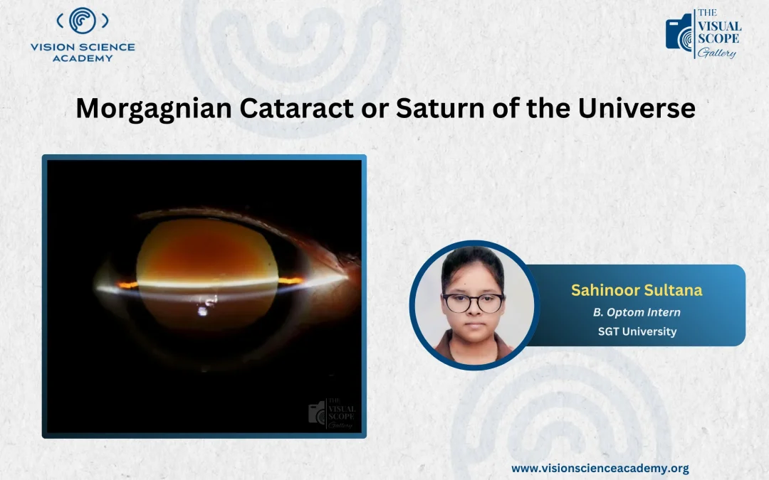
Sahinoor Sultana, B.Optom. Intern SGT University, Gurugram, India Figure: Slit Lamp Image Showing Morgagnian Cataract Image Summary A 59-years old male reported with a difficulty in seeing distance and near with the right eye, more compared...
by Vision Science Academy | Jan 1, 2026 | Photography
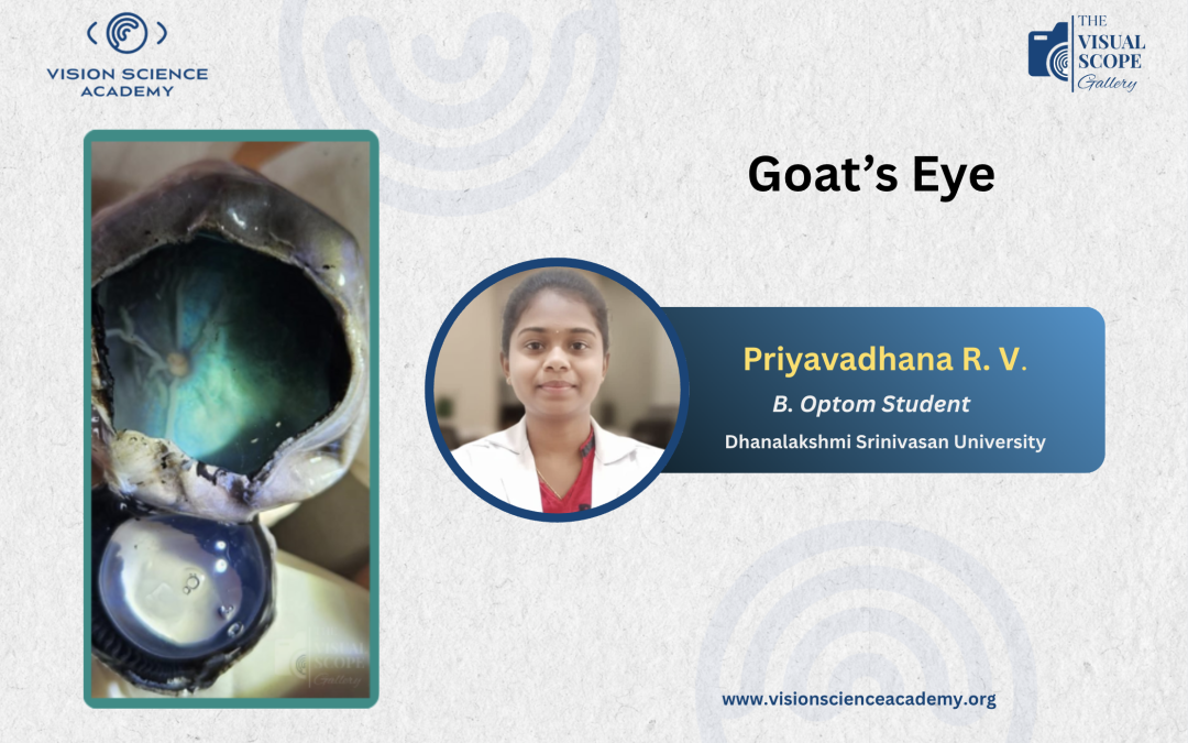
Priyavadhana R. V, B.Optom. Student Dhanalakshmi Srinivasan University, Trichy, India Figure: A Dissected View of Goat’s Eye Image Summary Goat eyes serve as a valuable research model in vision science, offering clear insights into eye...

by Vision Science Academy | Dec 1, 2025 | Photography
Haziel Rynjah, M. Optom. Assistant Professor, Royal Global University, Guwahati, India Figure: Slit-lamp Photograph of a Case with Multiple Superficial Punctate Keratitis (SPK) stained with Fluorescein Dye and Visualised in Cobalt Blue Light in a...
by Vision Science Academy | Nov 1, 2025 | Photography
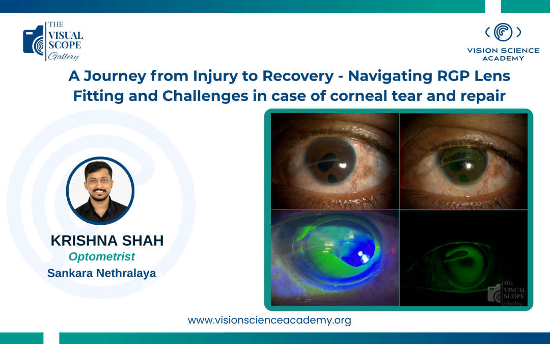
Krishna Shah, M. Optom. Optometrist, Sankara Nethralaya, Chennai, India Image: Rigid Gas Permeable Lens in Corneal Tear Repair Demonstrating Edge Lift This case follows a 24-year-old male who sustained a significant corneal injury from a wooden...
by Vision Science Academy | Oct 1, 2025 | Photography
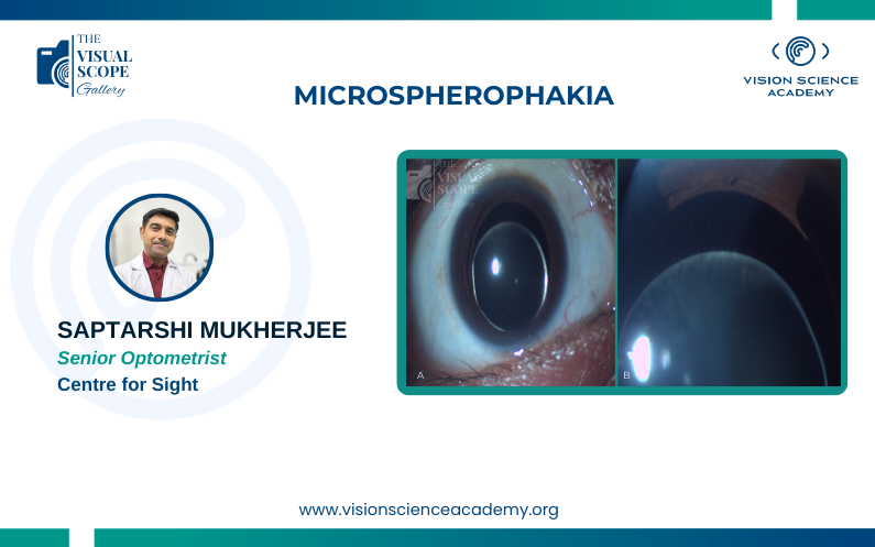
Saptarshi Mukherjee, M.Optom. Senior Optometrist, Centre for Sight, New Delhi, India Image: Slit lamp picture of Microspherophakia. A. Small Dislocated Crystalline Lens, B.Weak Zonules Image Summary Microspherophakia is a rare congenital...
by Vision Science Academy | Sep 1, 2025 | Photography
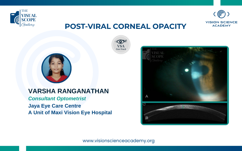
Varsha Ranganathan, B. Optom Clinical Optometrist, Jaya Eye Care Centre, A Unit of Maxi Vision Eye Hospital, Chennai, India Image: : A) Corneal Opacity as seen in the Slit Lamp Examination. B) Cross-sectional view of the cornea seen via Anterior Segment...
Recent Comments