by Vision Science Academy | Nov 1, 2025 | Photography
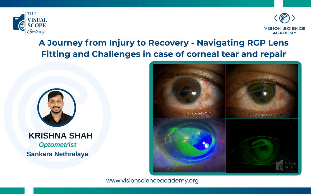
Krishna Shah, M. Optom. Optometrist, Sankara Nethralaya, Chennai, India Image: Rigid Gas Permeable Lens in Corneal Tear Repair Demonstrating Edge Lift This case follows a 24-year-old male who sustained a significant corneal injury from a wooden...
by Vision Science Academy | Oct 1, 2025 | Photography
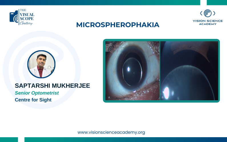
Saptarshi Mukherjee, M.Optom. Senior Optometrist, Centre for Sight, New Delhi, India Image: Slit lamp picture of Microspherophakia. A. Small Dislocated Crystalline Lens, B.Weak Zonules Image Summary Microspherophakia is a rare congenital...
by Vision Science Academy | Sep 1, 2025 | Photography
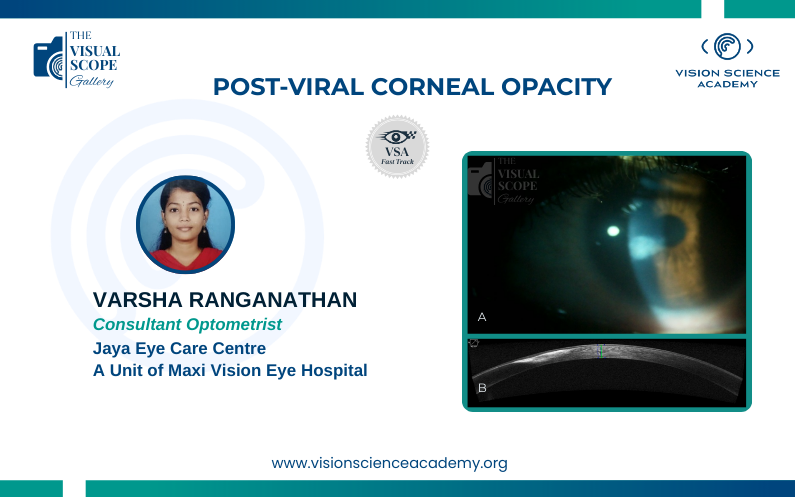
Varsha Ranganathan, B. Optom Clinical Optometrist, Jaya Eye Care Centre, A Unit of Maxi Vision Eye Hospital, Chennai, India Image: : A) Corneal Opacity as seen in the Slit Lamp Examination. B) Cross-sectional view of the cornea seen via Anterior Segment...

by Vision Science Academy | Aug 1, 2025 | Photography
Naman Kumar Shah, B.Optom. Clinical Optometrist, Dr. Patwardhan’s Nandadeep Eye Hospital, Sangli, India Image: Before and After Images of Metallic Foreign Body Removal Image Summary Before and after images of metallic foreign body...
by Vision Science Academy | Jul 1, 2025 | Photography
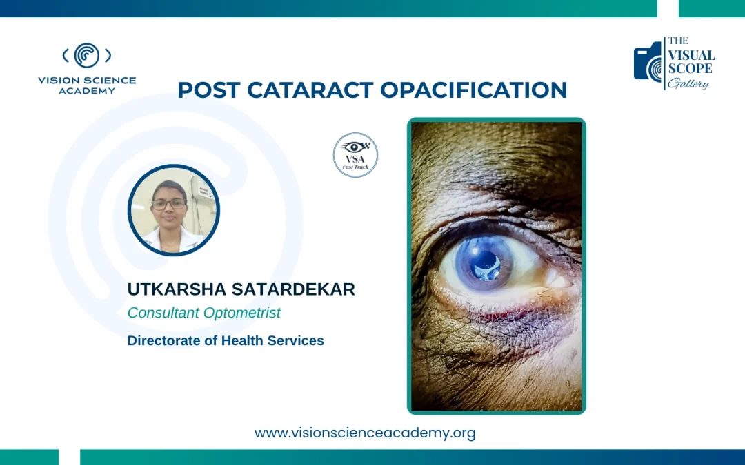
Utkarsha Satardekar, B. Optom Consultant Optometrist, Directorate of Health Services, Goa, India Image: Cloudy Intraocular Lens appearance; a sign of Post Cataract Opacification Image Summary The image is depicting an eye with post-cataract...
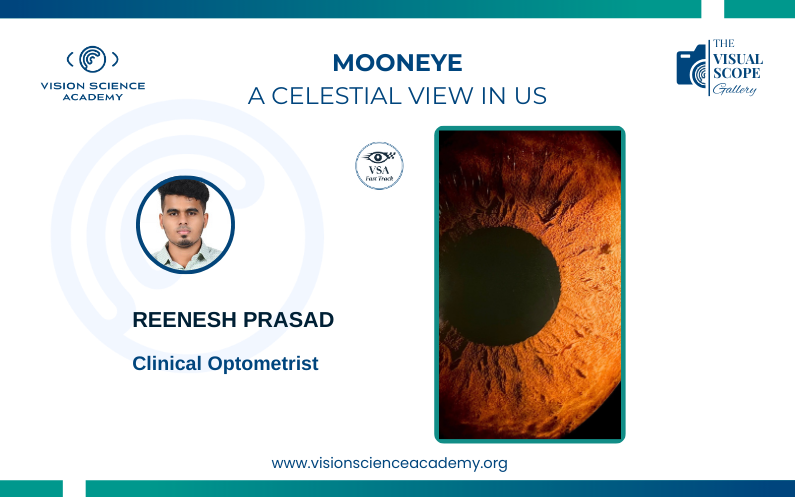
by Vision Science Academy | Jun 1, 2025 | Photography
Reenesh Prasad, Fellow in Clinical Optometry Clinical Optometrist, Chennai, India Image: Patterns of the Crypts of Iris Image Summary The intricate architecture of the iris crypts resembles the celestial topography of the moon, with its...

Recent Comments