
by Vision Science Academy | Aug 1, 2025 | Photography
Naman Kumar Shah, B.Optom. Clinical Optometrist, Dr. Patwardhan’s Nandadeep Eye Hospital, Sangli, India Image: Before and After Images of Metallic Foreign Body Removal Image Summary Before and after images of metallic foreign body...
by Vision Science Academy | Jul 1, 2025 | Photography
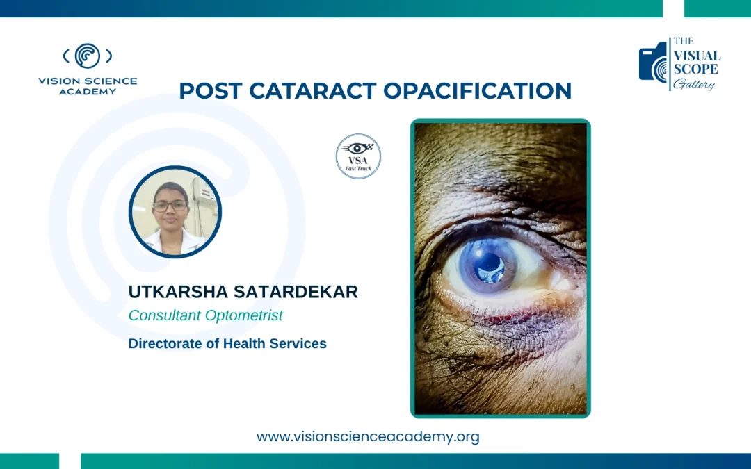
Utkarsha Satardekar, B. Optom Consultant Optometrist, Directorate of Health Services, Goa, India Image: Cloudy Intraocular Lens appearance; a sign of Post Cataract Opacification Image Summary The image is depicting an eye with post-cataract...
by Vision Science Academy | Jun 1, 2025 | Photography
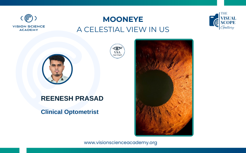
Reenesh Prasad, Fellow in Clinical Optometry Clinical Optometrist, Chennai, India Image: Patterns of the Crypts of Iris Image Summary The intricate architecture of the iris crypts resembles the celestial topography of the moon, with its...
by Vision Science Academy | Jun 1, 2025 | Photography
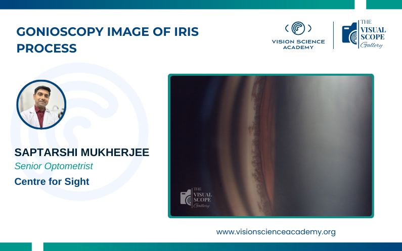
Saptarshi Mukherjee, M.Optom Senior Optometrist, Centre for Sight, New Delhi, India Image: Iris Process; thin lace like extended structure of iris tissue Image Summary Iris Process (IP) is fine extension of iris into posterior...
by Vision Science Academy | May 14, 2025 | Photography
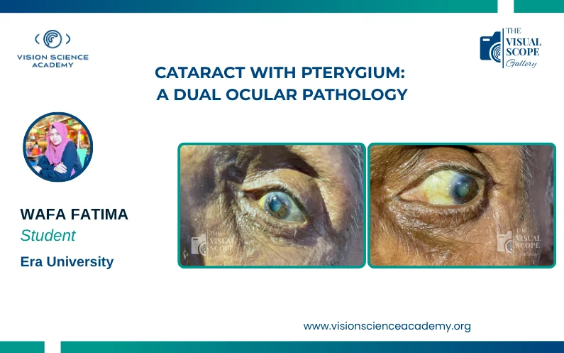
Wafa Fatima, B. Optom Student, Era University, Lucknow, India Image: Dual Pathology of Cataract and Pterygium in the eye Image Summary This image depicts an eye affected by both cataract and pterygium. The cloudy lens indicates...
by Vision Science Academy | May 14, 2025 | Photography
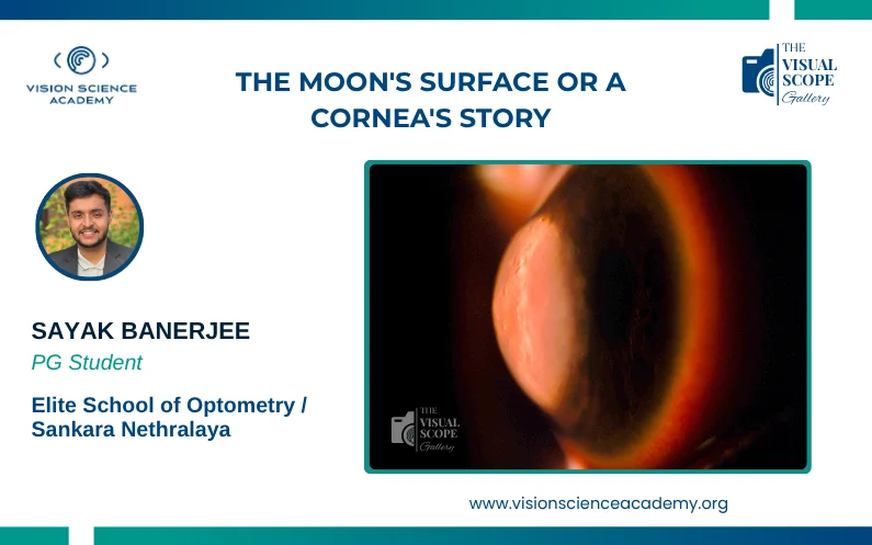
Sayak Banerjee, B. Optom PG Student, Elite School of Optometry/Sankara Nethralaya, Chennai, India Image: Sclerotic Scatter in a Keratoconic Eye with Hydrops Image Summary Captured with a Topcon SL D701 using the Sclerotic Scatter...
Recent Comments