


Riya Chatterjee, B. Optom
Consultant Optometrist, Lenskart, Bhubaneshwar, India
Neuromyelitis Optica Spectrum Disorders (NMOSD) are rare autoimmune conditions that primarily affect the optic nerves and spinal cord, leading to severe visual and neurological impairments. (1) Unlike Multiple Sclerosis (MS), NMOSD is characterised by the presence of anti-Aquaporin-4 (AQP4) antibodies, which target astrocytes in the central nervous system. (1) Early diagnosis is crucial to prevent irreversible damage, making the role of Optometrists essential in recognising early ocular manifestations and referring patients for prompt neurological evaluation.
Optometric Diagnosis of NMOSD
Optometrists play a pivotal role in detecting NMOSD-related visual symptoms, including sudden vision loss, optic neuritis, and pain with eye movement. The diagnostic process involves:
- Comprehensive Eye Examination: Patients with NMOSD often present with reduced visual acuity, Relative Afferent Pupillary Defect (RAPD), and optic disc swelling, which can be assessed using fundoscopy and Optical Coherence Tomography (OCT). (2)
- Visual Field Testing: NMOSD-related optic neuritis may cause central or altitudinal scotomas, requiring perimetry for early detection. (2)
- OCT Imaging: OCT helps in evaluating Retinal Nerve Fibre Layer (RNFL) thinning, a hallmark of optic neuritis progression. (2)
- Colour Vision Testing: Dyschromatopsia is an early sign of optic nerve involvement and can be assessed using Ishihara or Farnsworth D-15 tests. (2)
- Collaboration for Serological Testing: Since NMOSD is confirmed through AQP4 antibody testing, Optometrists must collaborate with Neurologists and Immunologists to facilitate serological analysis. (1)
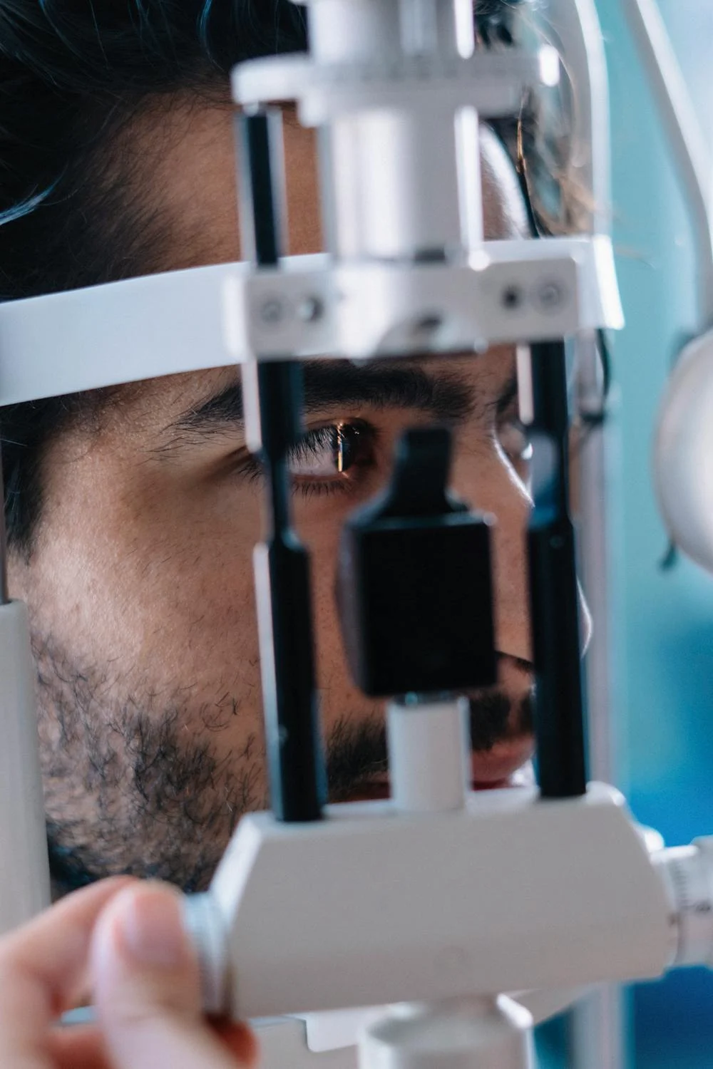

Figure 1: This image shows Retinal Examination being performed using slit lamp biomicroscopy.
Management Strategies for NMOSD
Although NMOSD has no definitive cure, Optometrists can assist in visual rehabilitation and management through:
- Corticosteroid Therapy Support: High-dose intravenous methylprednisolone is first-line treatment for acute attacks, reducing inflammation and preserving vision. (1)
- Low Vision Rehabilitation: Patients with persistent visual deficits benefit from magnifiers, contrast-enhancing aids, and adaptive technologies. (1)
- Prism Correction: Diplopia caused by optic neuritis or brainstem involvement can be managed using Fresnel prisms. (2)
- Ocular Surface Management: Chronic immunosuppressive therapy can cause dry eye disease, necessitating lubricants and punctal plugs. (2)
- Patient Education: Optometrists should educate patients on relapse indicators, such as sudden vision changes, and the need for immediate medical attention.
Emerging Insights in NMOSD
Recent studies have identified Myelin Oligodendrocyte Glycoprotein (MOG) antibodies as another potential biomarker in NMOSD patients who test negative for AQP4 antibodies. (3) Furthermore, advanced imaging techniques, including ultra-high-resolution OCT, now allow early detection of microstructural retinal changes, offering a non-invasive biomarker for disease monitoring. (2)
Conclusion
In summary, Optometrists play a vital frontline role in the early detection and ongoing management of NMOSD. Because optic neuritis is often the first presenting symptom, timely identification of reduced vision, RAPD, visual field defects, and OCT-detected RNFL thinning enables early referral for Aquaporin-4 Immunoglobulin G (AQP4-IgG) testing and neurological evaluation, which is crucial for preventing irreversible visual disability. (1) Additionally, Optometrists contribute to monitoring disease progression, recognising relapse indicators, and guiding patients through low-vision rehabilitation and ocular surface management. With the emergence of Myelin Oligodendrocyte Glycoprotein-Immunoglobulin G (MOG-IgG) testing, their role in identifying atypical optic neuritis has expanded further, strengthening interdisciplinary collaboration in diagnosis and treatment planning. (3)
References
- Mealy MA, Wingerchuk DM, Greenberg BM, Levy M. Neuromyelitis optica spectrum disorder: Diagnosis and management. Curr Opin Neurol. 2018;31(3):255–263.
- Bennett JL, de Seze J, Lana-Peixoto M, et al. Neuromyelitis optica and multiple sclerosis: Seeing differences through optical coherence tomography. Mult Scler. 2015;21(6):678–688.
- Jarius S, Paul F, Aktas O, et al. MOG encephalomyelitis: International recommendations on diagnosis and antibody testing. J Neuroinflammation. 2018;15(1):134.
About the Author


Riya Chatterjee
Consultant Optometrist