


Brajesh Kumar, M.Optom
Faculty Optometrist, Dr. Shroff’s Charity Eye Hospital, New Delhi
The retina is the innermost neurosensory layer of the eyeball and the only part of the human body where the blood vessels are directly visible. Retina is a mirror of many systemic diseases and its examination primarily helps to detect hypertension and diabetes. Examination of the retina is done by slit lamp biomicroscopy, direct ophthalmoscopy & indirect ophthalmoscopy. However, in order to document the findings a fundus drawing was earlier done followed by photos on film-based cameras. In today’s digital world the fastest way to document is through digital fundus photography.
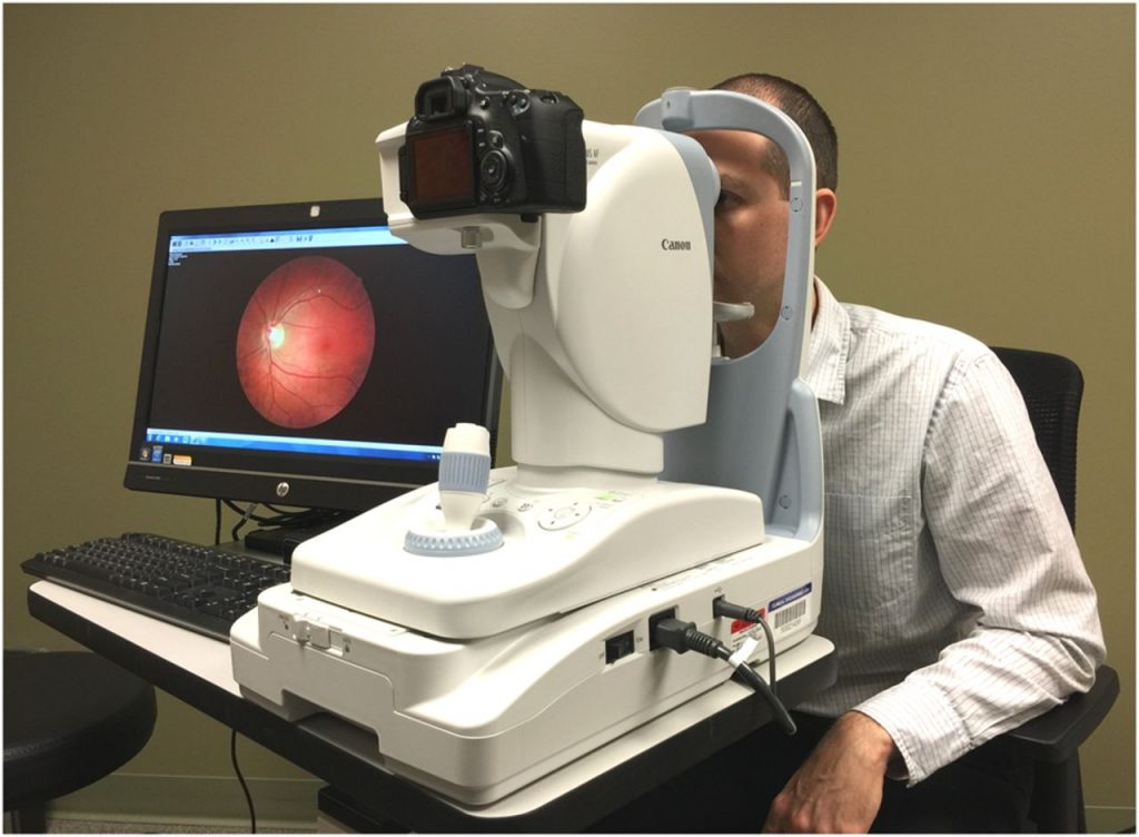
Digital fundus photography involves the process of taking photographs with the help of a fundus camera comprising of a microscope with filters. Recording the fundus images electronically, storing and manipulating them in a computer. The latest being a wide field camera providing documentation of the entire retina up to the periphery. A fundus photograph of a normal retina comprises of the optic disc, central retina known as macula, and retinal blood vessels (see Figure 1).


Figure 1: Colour fundus photo of the right eye showing the optic nerve head, the macula and the normal retinal vessels.
The advancing technology has helped to capture precise high-resolution fundus photos in a very short time, thereby, greatly expanding the horizon of ophthalmic photography.
Indications:
Diabetic retinopathy (DR): Various signs of DR1 are documented with fundus photos (Figure 2), often used in teleophthalmology.


Macular Pathologies: Macula is the most sensitive part of the retina primarily responsible for central vision. Various macular diseases such as Age-related Macular Degeneration (AMD), (see Figure 3) can be captured. 2


Figure 3: Colour fundus photo of the right eye showing a choroidal neovascular membrane with subretinal haemorrhage suggestive of AMD.
Glaucoma: It is a chronic disease of high intraocular pressure (IOP) damaging the optic nerve. Fundus photography plays a vital role in screening and documenting the eyes for glaucomatous changes of the optic nerve head 3,4 (see Figure 4).


Figure 4: Colour fundus photo of the right eye showing the cupping of the optic nerve head suggestive of glaucomatous damage.
Procedure:
- Check and clean the lens of the camera to avoid artifacts
- The patient details are entered to identify the images at a later date
- Explain the procedure to the patient and make him comfortable
- Adequate pupillary dilatation to achieve good quality images and to image the peripheral retina
- Patient is asked to keep his eyes open and maintain fixation on the given target.
- The patient is asked to look in different gaze which helps to acquire more of the peripheral retina in that quadrant
- Adequate flash light is required to avoid over exposure of images
- Final images are transferred and stored in the computer for later reference
Advantages:
- Portable (hand held cameras) and cost-effective
- Quick, safe, painless and non-invasive (no dye injection is required)
- Provides high resolution photographs
- Helps in documentation
- Helps in increasing reproducibility through standardized imaging protocols4
Disadvantages:
- Inadequate pupillary dilatation may lead to poor quality images
- Uncooperative patient or photophobia may lead to compromised image quality
- Increased external illumination may lead to involuntary blinking of eyes
To Summarise:
Digital Fundus Photography is an important tool of Ophthalmic photography which helps an Ophthalmologist in –
- Screening retinal pathologies
- Documentation of retinal and optic nerve pathologies
- Monitoring the progression of the disease
References
1. Mookiah, MR, Acharya UR, Fujita, H, Tan JH., Chua CK., Bhandary SV, Laude A, Tong L. Application of different imaging modalities for diagnosis of Diabetic Macular Edema: A review. Computers in biology and medicine 2015; 66:295–315.
2. Bernardes R, Serranho P, Lobo C: Digital Ocular Fundus Imaging: A Review. Ophthalmologica 2011;226:161-181.
3. Myers JS., Fudemberg SJ, Lee D. Evolution of optic nerve photography for glaucoma screening: a review. Clinical & experimental ophthalmology 2018;46(2): 169–176.
4. Abràmoff MD, Garvin, MK, Sonka M. Retinal imaging and image analysis. IEEE Rev Biomed Eng.2010 Jan 1; 3: 169–208.