


Niru Ranachand Kanchan, B. Optom Student,
Sri Manakula Vinayagar Medical College, Pondicherry, India
Dry Eye Disease (DED) is a multifactorial disease of the ocular surface that is caused by insufficient tear production and rapid tear evaporation, often due to age (menopause and post menopause), chronic environmental factors, etc. It significantly affects the Quality of Life (QoL) of a person. (1) Weakening symptoms of DED include dryness, redness, burning, and decreased vision; these affect the daily activities of the patient. A crucial impact of dry eye. Disease that results in impaired visual function. Most of the studies have found the prevalence of DED is between 5-50% of the population. (2) The female sex is a major risk factor for the development of DED, notably in post menopause women. This article explains how a hormonal disorder affects the ocular surface in a condition like DED with menopause in women.
Tear Film Anatomy
The anatomy and physiology of the tear film of the human eye is depicted in the image below.
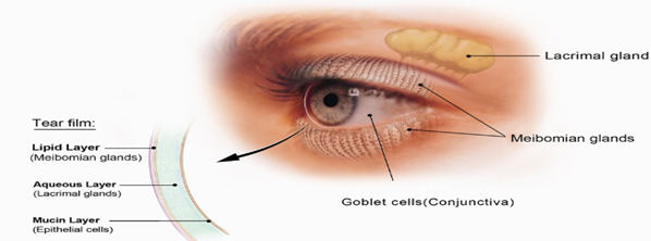

Figure 1: This image shows the layers of the tear film.
Image Courtesy:
https://www.linkedin.com/pulse/three-layers-tear-film-bridgitte-shen-lee-od
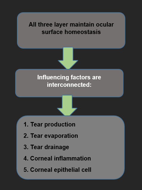

Figure 2: This flowchart explains ocular surface homeostasis.
Hormonal Effect on Dry Eye
The body cells and tissues of a human regulate the endocrine system. Hormones are constantly circulated in the ocular surface to expose their receptors. Endocrine anomalies are physiological and pathological conditions which cause DED. The physiological condition is menopause and morbid conditions are polycystic ovarian syndrome, androgen resistance. (3) They both condition the effects of both menopause and hormonal replacement therapy. So, the management of DED is hormonal therapeutics.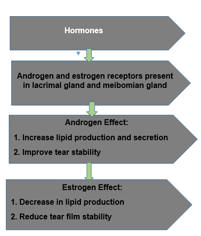

Figure 3. This flowchart explains the effects of androgen and estrogen in the tear film.
Androgen
Increases lacrimal gland production and immunoglobulin-A (IgA) secretion in androgen; it develops and progresses DED.
Estrogen and Progesterone
Both receptors are present on the ocular surface. Like conjunctiva, cornea, and glands such as the meibomian and lacrimal glands. As compared to androgen, estrogen indicates the antagonising effect on the meibomian gland. These effects of the estrogen on the meibomian gland that worsening of DED clinical features in the menopause on hormone replacement therapy.
Management of DED
Therapies are the successful treatment of DED. Such treatments are non-steroidal anti-inflammatory drugs such as corticosteroids and biological tear replacement, antibiotics, and tea tree oil are hormonal therapy. (4)
Anti-Inflammatory Therapy: NSAIDS
The Non-steroidal Anti-Inflammatory Drug (NSAIDs) mechanism blocks cyclooxygenase (COX) enzymes, and it inhibits prostaglandin production. The therapeutic effect of the NSAIDs is to reduce inflammation of the ocular surface, corneal sensitivity. It should be used only for short-term therapy; avoid long term use due to risk of corneal epithelial damage.
Hormonal Therapy: Anti-Androgen Therapy
Anti-androgen therapy is used in prostate cancer treatment. (4) It shows that the androgen deficiency is commonly associated with DED symptoms. The duration of anti-androgen therapy between is 3-96 months. It shows that in patients with elevation of ocular surface dryness and decreased Tear Breakup Time (TBUT), androgen plays a vital role in maintaining tear film stability. Post-menopausal women benefit from androgen supplementation. Transdermal androgen therapy is the best treatment for patients with androgen deficiency–related DED symptoms.
Conclusion
Sex hormone plays a vital role in the pathophysiology of DED in menopause and post menopause age group. It has the increased severity of DED symptoms and impairment in vision related function significantly reducing the overall QOL. In severe cases, anti-inflammatory therapy, hormonal replacement therapy is used and rarely surgical procedures are required.
References
- Peck, T., Olsakovsky, L., & Aggarwal, S. (2017). Dry eye syndrome in menopause and perimenopausal age group. Journal of mid-life health, 8(2), 51-54.
- Garcia-Alfaro, P., Garcia, S., Rodriguez, I., & Vergés, C. (2021). Dry eye disease symptoms and quality of life in perimenopausal and postmenopausal women. Climacteric, 24(3), 261-266.
- Gorimanipalli, B., Khamar, P., Sethu, S., & Shetty, R. (2023). Hormones and dry eye disease. Indian journal of ophthalmology, 71(4), 1276-1284.
- Nguyen, A., Kolluru, A., & Beglarian, T. (2023). Dry eye disease: A review of anti-inflammatory therapies. Taiwan Journal of Ophthalmology, 13(1), 3-12
About the Author


Niru Ranachand Kanchan
B. Optom Student,