


Gopi Krishnan G(1), Bhargavy S(2),
1M.Optom. Student, Dr. Agarwal’s Institute of Optometry, Chennai, India
2Assistant Professor Jr., Dr. Agarwal’s Institute of Optometry, Chennai, India
The phrase “Your eyes can’t lie” reflects a new truth in AI-powered healthcare. As diabetes silently affects millions, the need for early, non-invasive screening is urgent. The iris, the eye’s coloured part, has emerged as a promising diagnostic surface. This method combines ancient iridology with modern artificial intelligence (AI). By analysing iris patterns with AI, researchers aim to detect diabetes early, particularly in regions with limited access to diagnostics. (1)
Rethinking Iridology with AI
Iridology suggests that iris zones correspond to internal organs. Historically seen as alternative medicine, it is now being revisited using modern tools. Traditional maps link pancreatic function to specific iris areas. Advances in machine learning and image recognition now make it possible to validate these correlations through data. (2)
The lower nasal quadrant of the iris has been linked to the pancreas. (2) Subtle changes in texture or pigmentation in this region may suggest metabolic imbalance. AI algorithms trained on iris images are now identifying these patterns to help detect early signs of diabetes. (3)
Diabetes Detection: A Modern Challenge
Type 2 diabetes often develops unnoticed, delaying diagnosis and treatment. (4) In many areas, blood tests may be inaccessible or unaffordable. AI-driven iris scanning provides a contactless, scalable, and low-cost solution. (5) With undiagnosed diabetes on the rise globally, new screening technologies are vital to public health efforts. (6)
How the Technology Works
Several interventions have been proven effective in managing cognitive decline arising from visual impairment or chemotherapy:
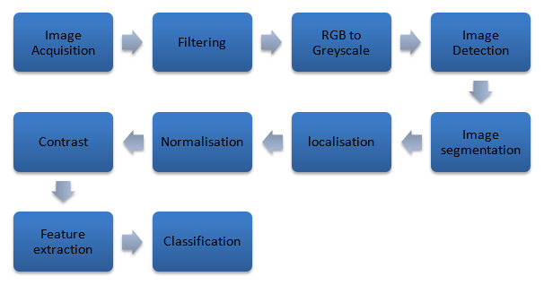

Figure 1: AI workflow diagram for iris analysis.
(Self-created by author)
Modern AI-based iris analysis involves a sophisticated, multi-step process from image capture to risk prediction:
1. Image Acquisition: A high-resolution iris image is captured using a digital camera, a smartphone with a macro lens, or a dedicated iris scanner. Proper lighting minimises reflections and shadows.
2. Pre-processing: The image undergoes enhancement steps, including brightness normalisation, edge detection, and noise reduction. This ensures consistency across different lighting and imaging conditions.
3. Iris Segmentation: Algorithms such as Hough Transform or Daugman’s rubber sheet model isolate the iris from the pupil, sclera, and eyelids. Accurate segmentation is critical for reliable feature extraction.
4. Feature Extraction: Convolutional Neural Networks (CNNs) analyse iris features such as crypt density, pigmentation variation, contraction furrows, and blood vessel visibility. These are studied especially in pancreas-related zones. (3)
5. Classification & Risk Scoring: AI models trained on labelled datasets (diabetic and non-diabetic) detect risk patterns and predict the likelihood of diabetes. Model accuracy improves through continuous learning.
6. Output Delivery & Integration: The result is displayed through clinical software or mobile apps. It can be linked with patient history for comprehensive risk assessment and telehealth referrals. (4)
The Future of Public Health Screening
Imagine clinics using handheld devices to scan your eye and deliver a diabetes risk score with no blood, no pain. This is becoming real as AI tools merge with mobile health platforms, making early screening possible in underserved regions. (9)
Conclusion
Blending iridology with deep learning offers a modern, non-invasive solution for diabetes screening. As technology advances, iris-based diagnostics could become a frontline tool combining ancient insight with AI-powered precision. (10)
References
- Azemin, M. Z. C., Hani, A. F. M., Huspi, S. H., Shakaff, A. Y. M., & Saad, F. S. A. (2018). Non-invasive diabetes detection using iris image analysis. Computer Methods and Programs in Biomedicine, 157, 153–162. https://doi.org/10.1016/j.cmpb.2018.01.004
- Jensen, B. (1982). The science and practice of iridology (2nd ed.). Bernard Jensen International. Retrieved from https://archive.org/details/sciencepracticeo0000bern
- Azemin, M. Z. C., Hani, A. F. M., Huspi, S. H., Shakaff, A. Y. M., & Saad, F. S. A. (2018). Non-invasive diabetes detection using iris image analysis. Computer Methods and Programs in Biomedicine, 157, 153–162. https://doi.org/10.1016/j.cmpb.2018.01.004
- Nasim, S., Akhtar, S., & Haider, S. (2022). Deep learning approach for early diabetes detection using iris image. Biomedical Signal Processing and Control, 76, 103670. https://doi.org/10.1016/j.bspc.2022.103670
- Kose, U., &Deperlioglu, O. (2021). Artificial intelligence-based medical diagnostic systems: Applications and trends. Procedia Computer Science, 196, 168–175. https://doi.org/10.1016/j.procs.2021.12.016
- Sheeladevi, S., Rani, P. K., & Ali, R. (2019). Vision screening for noncommunicable diseases: Expanding the role of eye care. Community Eye Health Journal, 32(105), 35–37.
- Bhaskaranand, M., Ramachandra, C., Bhat, S., Cuadros, J., &Nittala, M. G. (2016). Automated diabetic retinopathy screening and monitoring using retinal fundus image analysis. Journal of Diabetes Science and Technology, 10(2), 254–261. https://doi.org/10.1177/1932296816628546
- Hemanth, D. J., & Estrela, V. V. (2019). Deep learning for image processing applications in diabetic retinopathy detection. Journal of Medical Systems, 43, 289. https://doi.org/10.1007/s10916-019-1424-3
- Malandraki, G. A., & Georgiou, K. (2021). Artificial intelligence in clinical care: Current applications and future directions. Healthcare Technology Letters, 8(3), 53–58. https://doi.org/10.1049/htl2.12009
- Sharma, R., & Singh, A. (2020). Iris pattern recognition using deep learning techniques: A review. Artificial Intelligence Review, 53, 3975–4002. https://doi.org/10.1007/s10462-019-09780-2