


Sanjukta Jana, B. Optom.
Editorial Assistant Trainee, Vision Science Academy
In Vitro Fertilisation (IVF) is one of the most renowned Assisted Reproductive Technologies (ART) among people who are infertile. IVF involves the retrieval of mature oocytes, fertilisation of oocytes using sperm in the laboratory, and subsequent transfer of the zygote into the uterus, where it is implanted. (1)
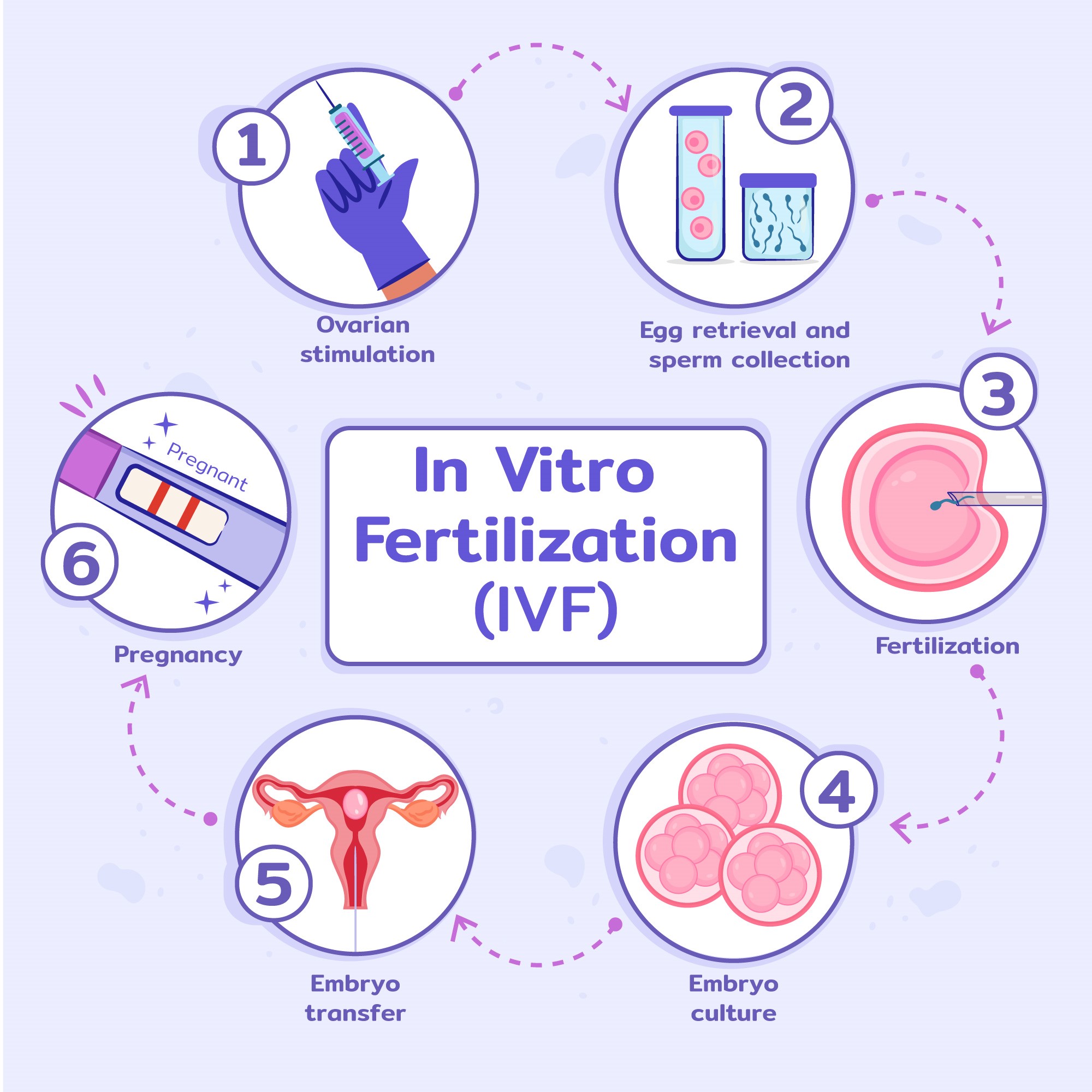

Figure 1: Pathophysiology of OSA
Increasing Global Use of ART
There has been a high usage of ART around the world. Some of the factors that have contributed to this rise include delayed childbearing, fertility loss due to lifestyle, increased access to reproductive care, and increasing acceptance of medical and elective fertility treatments in society. (1)
Rationale for Investigating Ocular and Developmental Effects After ART
The early weeks of pregnancy represent a period of great vulnerability to environmental, hormonal and metabolic interference. Early organ formation, including the development of eyes, occurs during this period. The nervous system tissues, neuroectodermal and mesodermal tissues, are highly coordinated in eye development, and the alterations in this environment can affect the process of ocular differentiation, formation of the optic nerve, or retinal development.(2)
Potential Influences Brought by ART
Hormonal Manipulation: Controlled ovarian hyperstimulation is one of the methods commonly employed in ART cycles to achieve supraphysiological levels of progesterone and oestrogen. (2, 3) These hormone levels are not similar to the early pregnancy natural endocrine profile and may be relevant in affecting cellular signalling pathways involving embryonic eye development. (4)
Embryo Manipulation and Epigenetic Regulation ART involves numerous embryo manipulation techniques, including prolonged culture, in-vitro fertilisation and micromanipulation techniques, including Intracytoplasmic Sperm Injection (ICSI). These interventions may affect epigenetic programming, including DNA methylation and genomic imprinting, that guide the developmental pathways of the early embryonic and ocular developmental stages. (5)
Perinatal and Neonatal Confounding Factors One of the characteristics of ART pregnancies is an increased prevalence of preterm births, the births with very low weights, and multiple pregnancies. Prematurity is a proven independent risk factor in the development of retinopathy of prematurity and refractive error, and with strabismus development (Slome et al., 2008). (5)
Literature Reporting No Additional Ocular Abnormalities in ART Children
Several studies have failed to establish a greater prevalence of ophthalmic abnormalities within the ART-conceived children as compared to their naturally conceived counterparts. Indicatively, a study that compared eye anomalies in children conceived through ICSI through a cohort study found that the procedure did not significantly increase the likelihood of refractive error, strabismus, or structural ocular anomalies. (6)
Epidemiological studies of large scale offer further information. A quasi-population study of ART-conceived children to the age of 18 years identified no regular trend of high ophthalmic morbidity. (7)
Even though certain subgroups did exhibit marginally higher risks, they were mostly reduced once confounding factors like prematurity, birth weight, socioeconomic status and parental traits were accounted. Such studies form the best evidence available as they have very large samples and thorough follow-up.
Systematic reviews that have produced the synthesis of ART and congenital anomaly information illustrate a great degree of heterogeneity in the outcomes documented and opposing outcomes in studies. (7,8)
Evidence Reporting Ocular and Visual Abnormalities in ART -Conceived Children
Several early investigations suggested higher rates of ocular abnormalities among ART-conceived children. For example, Anteby et al. reported increased frequencies of refractive errors, strabismus, congenital cataract, optic nerve anomalies, and colobomas in IVF-born children. (2) These abnormalities, while relatively uncommon, raised concerns regarding potential developmental disruption during embryogenesis.
Other studies have highlighted functional visual differences. Wennerstrom et al. observed delayed visual maturation and atypical fixation patterns among ART infants when compared with naturally conceived controls. (7) These findings suggest that neuro-visual development may be subtly altered in some ART-conceived infants.
Across studies reporting positive associations, observed abnormalities include refractive errors, anisometropia, strabismus or misalignment, congenital structural anomalies, optic nerve defects, and delayed visual maturation. However, these findings are inconsistent across cohorts and often limited by small sample sizes.
| Section | Source | Key Findings |
|---|---|---|
| Observational Studies Showing Ocular Differences | Anteby et al. |
Higher frequency of refractive errors in IVF-conceived children. Increased strabismus compared with naturally conceived peers. Reported cases of congenital cataract. Optic nerve anomalies documented. Presence of colobomas in some ART children. |
| Functional Visual Differences Reported in ART Infants | Wennerström et al. |
Delayed visual maturation in ART infants. Atypical fixation patterns observed. Visual responses less mature than naturally conceived controls. |
Table 1: Key Findings from various studies summarised
Confounding Factors and Methodological Issue
Perinatal and Neonatal Confounders
Ocular development has been reported to be affected by preterm, low birth weight, oxygen, and Neonatal Intensive Care Unit (NICU) exposure. (6) These considerations are more common in ART pregnancies, and they could be the cause of certain ocular abnormalities.
Parental, Genetic and Infertility-Related Factors
Infertility may have underlying causes, such as the age of the parents or genetic predispositions, which may cause foetal development without ART. (5)
ART Protocol variability
Stimulation differences in regimens, embryo manipulation, culture media, ICSI use, and fresh versus frozen transfer led to variability, which makes differentiation of outcomes difficult.
Study Design Limitations
Small sample sizes, lack of consistency in the ophthalmic evaluation, and uneven diagnostic criteria among the studies make comparisons difficult and undermine the evidence base.
Clinical Implications of Eye Care
Need for Early Screening: Considering the mixed evidence, periodical and early visual examination of children born with ART should be used.
Parental Counselling: The combination of communication will be necessary to avoid inappropriate panic and focus on the timely eye check-ups.
Collaborative Care: The collaboration between Optometrists, Paediatricians, Ophthalmologists, and reproductive specialists should be established to check the development of vision.
Research Recommendations: Subsequent research needs to have standard ophthalmic procedures, ART procedural reporting and longitudinal follow-up.
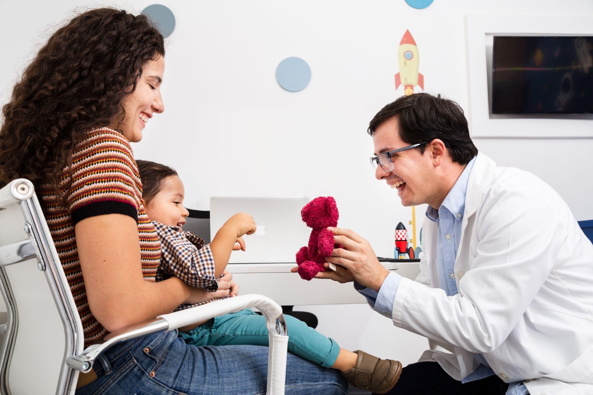

Figure 2: Play based interaction with parental involvement. Along with emphasising the birth history of child , conception history can also be included as a probe during pediatric history taking
Conclusion
The studies on the correlation of ART and ocular results are inconclusive. There are studies of higher prevalence rates of ocular abnormalities and others of normal developmental patterns. It is difficult to interpret due to confounding issues, methodological shortcomings, and variability of ART. Although ART is safe and viable as a fertility technology, infants conceived by ART should be screened in terms of vision shortly after birth. The need to have quality longitudinal studies would help shed light on the issue of whether ART is a limiting factor in ocular development risk.
References
- Zaher, N., & Halawa, A. (2023). In vitro fertilisation. StatPearls Publishing.
- Anteby, I., Cohen, E., Anteby, E., & BenEzra, D. (2001). Ocular manifestations in children born after in vitro fertilization. Archives of Ophthalmology, 119(10), 1525–1529.
- Wikstrand, M. H., Strömland, K., Flodin, S., Bergh, C., Wennerholm, U.-B., & Hellström, A. (2006). Ophthalmological findings in children born after intracytoplasmic sperm injection. Acta Ophthalmologica Scandinavica, 84(2), 177–181.
- De Gennaro, L., & Bucci, S. (2018). Assisted reproductive technologies and ocular development: A systematic review. Journal of Pediatric Ophthalmology and Strabismus, 55(5), 318–326.
- Sutcliffe, A. G., & Ludwig, M. (2007). Outcome of assisted reproduction. The Lancet, 370(9584), 351–359.
- Holmström, G., Larsson, E., & Hellström, A. (2014). Eye problems in children born prematurely. Acta Paediatrica, 103(1), 61–66.
- Wennerstrom, K., Nordström, M., & Sabel, K. G. (2002). Visual function in infants born after assisted reproductive techniques. Acta Ophthalmologica Scandinavica, 80(2), 137–142.
About the Author


Sanjukta Jana is a Postgraduate Optometry Student at SRM Medical College Hospital and Research Centre, with a focused research interest in Myopia, particularly the protective role of outdoor exposure and the association between light environment and Myopia development. She has a strong passion for scientific and academic writing and has been an active contributor to the Vision Science Academy, where her work has been widely recognised. She has achieved top positions in multiple academic competitions and has been awarded Visionary Blogger of the Month on several occasions across Vision Science Academy platforms. She aims to bridge clinical research and knowledge dissemination to support evidence-based vision care.