


Avantika Bind, B. Optom.
M. Optom Student, The Sankara Nethralaya Academy, Chennai, India
The cornea and conjunctiva form the outer surface of the eye, protected by a tear film from environmental factors. The transparent cornea, which lacks blood vessels, acts as a barrier and contributes to about 75% to the eye’s focusing ability. Its epithelium, made of non-keratinised stratified squamous cells with tight junctions, protects the eye and provides a smooth surface for vision, regenerating every 7-10 days through limbal stem cell differentiation. (1-3)
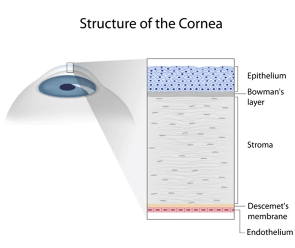

Figure 1: Layers of the cornea
Persistent Epithelial Defects: Causes and Conventional Treatments
A healthy epithelium is essential for protecting the eye from infection and preventing damage to underlying tissues. Acute epithelial defects in a healthy cornea typically heal within 7-14 days. (4,5) Persistent epithelial defects (PEDs) occur when epithelialisation fails, causing discomfort, vision issues, and potential disruption due to adhesion problems, stem cell deficiency, trauma, medications, or infections. (6)
Treatment for PEDs typically starts with conservative methods such as preservative-free lubricating eye drops, bandage contact lenses, and punctal plugs. (7) If these are ineffective, options include autologous serum eye drops and surgical procedures like debridement and amniotic membrane grafts, with autologous serum and platelet-rich plasma drops as second-line treatments. (8-10) If these fail, more aggressive options like amniotic membrane transplantation or surgery may be necessary. Emerging treatments, such as recombinant nerve growth factor and epidermal growth factor, provide non-invasive alternatives, while corneal neurotisation shows promise in restoring sensation and enhancing healing. (11,12)
Emerging Role of Insulin in Corneal Healing
Insulin-like Growth Factors (IGFs) are crucial for the growth and proliferation of corneal epithelial cells through insulin and IGF receptors. (13,14) Insulin promotes the migration of human epidermal keratinocytes and supports wound healing. It is present on the corneal surface and in the tear film, enhancing epithelial cell growth and potentially aiding in resistant epithelial defects. (15-17)
Topical insulin is safe for ocular use in humans and shows promise for treating Persistent Epithelial Defects. (18-20) Insulin, a peptide closely related to Insulin-like Growth Factor (IGF), stimulates keratinocyte migration and plays a role in wound healing. (16,21) The importance of glucose for corneal cell functionality is known, with glucose uptake occurring independently of insulin via the active glucose transporter GLUT1, which increases after wounding to support cell migration and proliferation. (22,23)
The mechanism by which insulin aids epithelialisation is still unclear. However, in diabetic animals, topical insulin has been shown to normalise DNA synthesis in basal epithelial cells within 48 hours post-wound, suggesting a role in cell proliferation for re-epithelialisation. Additionally, insulin may help regulate receptor homeostasis in corneal epithelial cells. (13)
A study concluded that topical insulin 1 unit, four times daily, improved corneal epithelial healing in diabetic patients post-vitrectomy, with no adverse events. (24) It was found that topical insulin eye drops (100 units/mL, twice daily) caused no ocular side effects in healthy individuals, and no systemic absorption was observed; the drops were as comfortable and clinically harmless as sterile saline. (25,26)
Conclusion
Topical insulin is safe for ocular use and effectively stimulates corneal re-epithelisation in persistent PEDs. By promoting cell growth, reducing inflammation, and aiding tissue repair, it provides a valuable strategy for managing challenging cases. Its therapeutic potential in epithelial healing is gaining recognition.
References
- Eghrari, A.O.; Riazuddin, S.A.; Gottsch, J.D. Overview of the Cornea: Structure, Function, and Development. Prog. Mol. Biol. Transl. Sci. 2015, 134, 7–23.
- Lu, L.; Reinach, P.S.; Kao, W.W. Corneal epithelial wound healing. Exp. Biol. Med. 2001, 226, 653–664
- Dhillon, H.K.; Bahadur, H.; Raj, A. A comparative study of tarsorrhaphy and amniotic membrane transplantation in the healing of persistent corneal epithelial defects. Indian. J. Ophthalmol. 2020, 68, 29–33.
- Vaidyanathan U, Hopping GC, Liu HY, et al. Persistent corneal epithelial defects: a review article. Med Hypothesis Discov Innov Ophthalmol J 2019; 8(3): 163–176.
- Ziaei M, Greene C and Green CR. Wound healing in the eye: therapeutic prospects. Adv Drug Deliv Rev 2018; 126: 162–176.
- Diaz-Valle D, Burgos-Blasco B, Gegundez-Fernandez JA, Garcia-Caride S, Puebla-Garcia V, Peña-Urbina P, Benitez-del-Castillo JM. Topical insulin for refractory persistent corneal epithelial defects. European Journal of Ophthalmology. 2021 Sep;31(5):2280-6.
- Katzman, L.R.; Jeng, B.H. Management strategies for persistent epithelial defects of the cornea. Saudi J. Ophthalmol. 2014, 28, 168–172.
- López-Plandolit S, Morales MC, Freire V, et al. Plasma rich in growth factors as a therapeutic agent for persistent cor neal epithelial defects. Cornea 2010; 29(8): 843–848.
- Tsubota K, Goto E, Shimmura S, et al. Treatment of persis tent corneal epithelial defect by autologous serum applica tion. Ophthalmology 1999; 106(10): 1984–1989.
- Rosenthal P, Cotter JM and Baum J. Treatment of persistent corneal epithelial defect with extended wear of a fluid-ven tilated gas-permeable scleral contact lens. Am J Ophthalmol 2000; 130(1): 33–41.
- Pflugfelder SC, Massaro-Giordano M, Perez VL, et al. Topical recombinant human nerve growth factor (Cenegermin) for neurotrophic keratopathy: a multicenter randomized vehicle-controlled pivotal trial. Ophthalmology 2019; 127(1): 14–26.
- Moon HS, Li L, Yoon HJ, et al. Effect of epidermal growth factor ointment on persistent epithelial defects of the cornea. BMC Ophthalmol 2020; 20(1): 147.
- Titone R, Zhu M and Robertson DM. Insulin mediates de novo nuclear accumulation of the IGF-1/insulin hybrid receptor in corneal epithelial cells. Sci Rep 2018; 8(1): 4378.
- Trosan P, Javorkova E, Zajicova A, et al. The supportive role of insulin-like growth factor-I in the differentiation of murine mesenchymal stem cells into corneal-like cells. Stem Cells Dev 2016; 25(11): 874–881.
- Shanley LJ, McCaig CD, Forrester JV, et al. Insulin, not leptin, promotes in vitro cell migration to heal monolayer wounds in human corneal epithelium. Invest Ophthalmol Vis Sci. 2005; 45:1088‒1094
- Naeser P. Insulin receptors in human ocular tissues: immunohistochemical demonstration in normal and diabetic eyes. Ups J Med Sci. 1997;102:35‒40
- Trosan P, Javorkova E, Zajicova A, et al. The supportive role of insulin-like growth factor-I in the differentiation of murine mesenchymal stem cells into corneal-like cells. Stem Cells Dev 2016; 25(11): 874–881.
- Fai S, Ahem A, Mustapha M, et al. Randomised controlled trial of topical insulin for healing corneal epithelial defects induced during vitreoretinal surgery in diabetics. Asia Pacific J Ophthalmol 2017; 6(5): 418–424
- Diaz-Valle, D.; Burgos-Blasco, B.; Gegundez-Fernandez, J.A.; Garcia-Caride, S.; Puebla-Garcia, V.; Peña-Urbina, P.; Benitez-Del Castillo, J.M. Topical insulin for refractory persistent corneal epithelial defects. Eur. J. Ophthalmol. 2021, 31, 2280–2286.
- Diaz-Valle, D.; Burgos-Blasco, B.; Rego-Lorca, D.; Puebla-Garcia, V.; Perez-Garcia, P.; Benitez-Del-Castillo, J.M.; Herrero-Vanrell, R.; Vicario-de-la-Torre, M.; Gegundez-Fernandez, J.A. Comparison of the efficacy of topical insulin with autologous serum eye drops in persistent epithelial defects of the cornea. Acta Ophthalmol. 2022, 100, e912–e919.
- Shanley, L.J.; McCaig, C.D.; Forrester, J.V.; Zhao, M. Insulin, not leptin, promotes in vitro cell migration to heal monolayer wounds in human corneal epithelium. Investig. Ophthalmol. Vis. Sci. 2004, 45, 1088–1094.
- Rocha, E.M.; Cunha, D.A.; Carneiro, E.M.; Boschero, A.C.; Saad, M.J.; Velloso, L.A. Identification of insulin in the tear film and insulin receptor and IGF-1 receptor on the human ocular surface. Investig. Ophthalmol. Vis. Sci. 2002, 43, 963–967.
- Stuard, W.L.; Titone, R.; Robertson, D.M. The IGF/Insulin-IGFBP Axis in Corneal Development, Wound Healing, and Disease. Front. Endocrinol. 2020, 11, 24. [CrossRef] [PubMed]
- Bastion MLC, Ling KP. Topical insulin for healing of diabetic epithelial defects? a retrospective review of corneal debridement during vitreoretinal surgery in Malaysian patients. Med J Mal. 2013; 68:207‒215.
- Bartlett JD, Turner-Henson A, Atchison JA, et al. Insulin administration to the eyes of normoglycemic human volunteers. J Ocul Pharmacol Ther. 2009; 10:683‒690.
- Bartlett JD, Slusser TG, Turner-Henson A, et al. Toxicity of insulin administration chronically to human eye in vivo. J Ocul Pharmacol Ther. 2009; 10:101‒107.