Anshi Jha,
B.Optom Student, Uttar Pradesh University of Medical Sciences, Etawah, India
Can the Eye Reveal Disease Long Before Symptoms Appear?
Imagine a young patient who comes in for a routine eye check-up. Vision is clear, retina looks healthy, and yet, hidden deep within the microvasculature, silent changes may already be unfolding. This is where Optical Coherence Tomography Angiography (OCTA) is rewriting the story of early diagnosis.
Often described as a “window to the body”, the retina offers far more than a view of ocular health. OCTA, a non-invasive and dye-free imaging technique, allows us to capture these subtle vascular changes, long before they turn into visible disease. (1)
Seeing Beyond Structure: How OCTA Works?
Traditional OCT shows us the layers of the retina. OCTA takes it further by detecting the movement of red blood cells to create detailed vascular maps. (2) With this technology, we can visualise blood flow in the superficial and deep capillary plexus and even the choriocapillaris, all without injecting dyes.
In simple terms, OCTA lets us go beyond anatomy to observe function, giving us a chance to detect disease in its earliest stage.
Silent Warnings: Diabetes and Hypertension
Both Diabetes and Hypertension are notorious for causing retinal damage, but often too late by the time it becomes clinically visible. OCTA helps uncover early warning signs:
1. Microvascular Dropout
Tiny non-perfusion areas seen in pre-diabetic patients, even when the fundus looks normal. (2)
2. Reduced Capillary Density
Subtle vascular compromise linked with hypertension. (3)
3. Vessel Tortuosity
Irregular blood vessel patterns that hint at progression towards retinopathy.
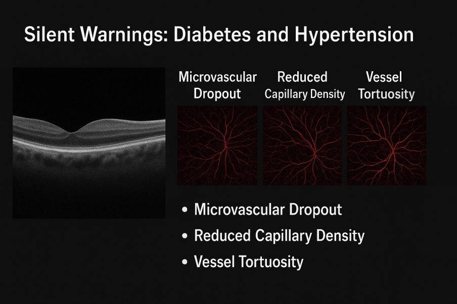
Figure 1: Created by Author
For an optometrist, this means we can step in earlier, guiding patients toward systemic care before sight is threatened
Beyond the Eye: Glaucoma and Neuro-degeneration
The scope of OCTA does not end with vascular diseases.
In glaucoma, OCTA has shown reduced peripapillary vessel density that may appear even before visual field loss. (3)
In Alzheimer’s and Parkinson’s Disease, studies suggest retinal microvascular alterations mirror what is happening in the brain (3)
This transforms the role of the optometrist, from being the guardian of sight to potentially being an early screener for neurological and systemic disorders.
Research Highlights Worth Noting
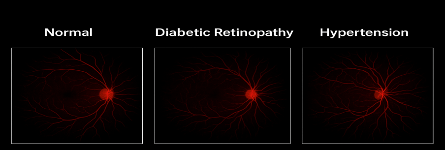
Figure 2: OCTA Showing Reduced Vessel Density in Diabetic Retinopathy and Hypertension compared to Healthy Eye
Recent investigations have shed light on OCTA’s broader potential:
- Diabetes: Pre-diabetic patients already show reduced vascular density, confirming OCTA as a tool for early intervention. (2)
- Hypertension: OCTA detects vascular irregularities well before clinical fundus changes. (3)
- Neuro-degeneration: Pilot studies suggest Alzheimer’s patients exhibit distinct OCTA patterns compared to age-matched healthy controls. (4)
Why Optometrists Matter in This Story
With OCTA, optometrists are uniquely positioned to detect what others might miss:
- Catching “invisible” preclinical changes that ophthalmoscopy or fundus photography cannot.
- Offering a non-invasive biomarker for systemic health.
- Collaborating with physicians and neurologists for holistic patient care.
The challenges such as cost, training, and image interpretation are real. However, the benefits make OCTA a technology that optometry cannot afford to ignore. (1)
Looking Ahead: The Future of Predictive Eye Care
The next chapter in OCTA involves Artificial Intelligence. Algorithms that can flag early risks automatically may soon assist clinicians, while portable OCTA devices promise to bring advanced diagnostics to under-served communities.
In the near future, optometrists may not only diagnose disease but predict it, redefining their role in preventive healthcare.
Conclusion
OCTA is more than just a new imaging modality. It represents a paradigm shift, a tool that allows us to see disease before it strikes. For optometrists, it opens a chance to protect vision and overall health by detecting silent systemic risks. The retina, once thought to reflect only the eye, is proving to be a mirror of the body itself.
References
- Spaide RF, Klancnik JM, Cooney MJ. Retinal vascular layers imaged by fluorescein angiography and OCT angiography. JAMA Ophthalmol. 2015.
- Carnevali A, et al. Optical coherence tomography angiography: A useful tool for diagnosis of diabetic retinopathy. Retina. 2017.
- Yarmohammadi A, et al. Vessel density in glaucoma measured with OCT angiography. Ophthalmology. 2016.
- Jiang H, et al. Retinal microvascular networks in Alzheimer’s disease: OCTA study. Invest Ophthalmol Vis Sci. 2018.
Recent Comments