


Rasmiya Bakrutheen, B. Optom Student
Sri Manakula Vinayagar Medical College and Hospital, Pondicherry, India
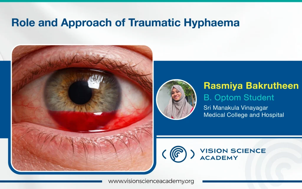
Traumatic Hyphaema is defined as the accumulation of blood within the anterior chamber of the eye following ocular trauma, most commonly blunt injury. It represents a significant cause of ocular morbidity worldwide, particularly among children and young adults engaged in sports and high-risk activities. Although many cases resolve without permanent visual impairment. Traumatic Hyphaema may be associated with serious complications such as secondary haemorrhage, elevated Intraocular Pressure (IOP), corneal blood staining, optic nerve damage, and late-onset Glaucoma. (1) Effective management requires careful evaluation, close monitoring, and timely intervention to minimise short- and long-term sequelae.
Pathophysiology
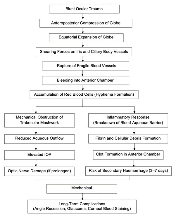

Image 1: This image shows the pathophysiology of blunt ocular trauma
Clinical Evaluation
Initial evaluation should include a detailed history of the injury, including mechanism, timing, and use of anticoagulants or antiplatelet agents.
Ocular examination includes assessment of visual acuity, pupillary reactions, slit-lamp biomicroscope to grade Hyphaema, and IOP measurement using applanation tonometry when feasible. Hyphaema is commonly classified into four grades based on the volume of blood in the anterior chamber. (2)
Conservative (Medical) Management
Most traumatic Hyphema are treated conservatively with bed rest, head elevation (30– 45°), activity restriction, and eye shielding. Topical corticosteroids reduce inflammation, and cycloplegics relieve pain and prevent synechiae. Aspirin and NSAIDs are avoided. Elevated IOP is managed with topical aqueous suppressants; systemic carbonic anhydrase inhibitors should be avoided in sickle cell disease. Antifibrinolytics may reduce secondary haemorrhage, but their routine use remains controversial. (2)
Surgical Management
Surgical intervention is reserved for selected cases where medical therapy fails or complications arise. Indications include persistent total Hyphaema, uncontrolled elevated IOP despite maximal medical therapy, corneal blood staining, or recurrent secondary haemorrhage. Anterior chamber washout is the most performed procedure and aims to remove accumulated blood and restore aqueous outflow.
Prognosis and Follow-Up
Prognosis is generally good in mild cases. Outcomes depend on injury severity and associated ocular damage. Long-term follow-up is essential to detect complications such as angle recession Glaucoma.
Conclusion
Traumatic Hyphaema is a potentially vision-threatening condition requiring prompt diagnosis and individualised management. While most cases resolve with conservative treatment, careful monitoring is essential to prevent complications. Special populations, such as patients with sickle cell disease, require modified management strategies. A structured, evidence-based approach and long-term follow-up are key to achieving optimal visual outcomes.
References
- Wilson II, F. M. (1980). Traumatic hyphema: pathogenesis and management. Ophthalmology, 87(9), 910-919.
- Walton, W., Von Hagen, S., Grigorian, R., & Zarbin, M. (2002). Management of traumatic hyphema. Survey of ophthalmology, 47(4), 297-334.
- Read, J., & Goldberg, M. F. (1974). Comparison of medical treatment for traumatic hyphema. Transactions of the American Academy of Ophthalmology and Otolaryngology, 78(5), OP799-OP815.
- Edwards, W. C., & Layden, W. E. (1973). Traumatic hyphema: a report of 184 consecutive cases. American journal of ophthalmology, 75(1), 110-116.
- Spoor, T. C., Kwitko, G. M., O’Grady, J. M., & Ramocki, J. M. (1990). Traumatic hyphema in an urban population. American journal of ophthalmology, 109(1), 23-27.
- American Academy of Ophthalmology. Basic and Clinical Science Course: Section 7 – Orbit, Eyelids, and Lacrimal System. AAO; latest edition.
About the Author


Rasmiya Bakrutheen
Optom Student,