


Rinsha Sadiqali
B. Optom Student, IIVM College of Optometry, Coimbatore, India
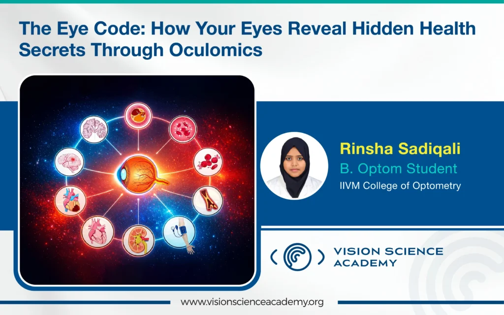
The retina is often called the ‘window to the brain’ because it shares a common embryological origin with neural tissue, has a rich neurovascular structure, and can be imaged non-invasively. Even subtle retinal changes can signal neurological or systemic diseases. More broadly, the eye serves as a ‘unique window to health,’ as many systemic disorders manifest ocular features across different layers of the eye. The term “oculome” refers to this full range of ocular features that reflect systemic health and disease, and the systematic analysis of these features through multimodal imaging to identify systemic biomarkers is known as Oculomics. (1)
How Do the Oculomics Work?
Oculomics starts by identifying ophthalmic biomarkers and using advanced imaging methods such as Electroretinogram (ERG), Optical Coherence Tomography (OCT), Confocal Scanning Laser Ophthalmoscopy (CSLO), Fluorescence Lifetime Imaging Ophthalmoscopy (FLIO), and Optical Coherence Tomography Angiography (OCT-A) to capture high-resolution structural and functional data from the eye. These imaging techniques generate large datasets that, when analysed with Artificial Intelligence (AI) and machine learning algorithms, help detect patterns, extract disease-specific biomarkers, and predict disease onset or progression.
Oculomics has demonstrated significant potential in linking ocular biomarkers to neurodegenerative diseases such as Alzheimer’s disease, Parkinson’s disease, Amyotrophic Lateral Sclerosis (ALS), Huntington’s disease, and other diseases, as described in Table 1. With advanced tools such as eye-tracking and next-generation imaging, diagnostic accuracy continues to improve. Overall, Oculomics enables earlier detection, better monitoring, and timely intervention for systemic diseases, enhancing patient quality of life. (2)


Figure 1: This Image shows the workflow of Oculomics.
Image Courtesy: Created by the author
Systemic Diseases and Their Ocular Biomarkers Systemic Diseases and Their Ocular Biomarkers
| Systemic Disease | Key Ocular Biomarkers |
|---|---|
| Multiple Sclerosis |
Retinal Nerve Fiber Layer (RNFL) thinning, optic nerve head changes |
|
Parkinson’s Disease, Alzheimer’s Disease |
Reduced retinal dopamine signals, thinning of inner retinal layers |
| Idiopathic Intracranial Hypertension | Papilledema, visual field defects |
| Schizophrenia |
Abnormal ERG responses, inner retinal layer thinning. (3) |
| Sarcopenia |
Blepharoptosis (drooping eyelid), decreased levator muscle function. (4) |
| Aneurysm Risk |
Retinal Vascular Features (RVFs), Retinal vessel tortuosity, Abnormal vascular branching patterns, Microvascular structural changes visible on retinal imaging. (5) |
| Rheumatoid Arthritis | Retinal vasculitis, uveitis |
|
Coronary Artery Disease / Cardio Vascular Diseases (CVD) Risk |
Arteriolar narrowing, venular widening |
| Chronic Kidney Disease |
Arteriolar narrowing, microvascular rarefaction |
Table 1: This table highlights the summary of systemic disease and ocular biomarkers.
What is the Role of Artificial Intelligence?
AI plays a transformative role in oculomics by enabling automated detection of subtle retinal biomarkers linked to systemic diseases. Using deep-learning analysis of OCT, OCTA, and fundus images, AI models can non-invasively predict conditions such as coronary artery disease, ischemic stroke, myocardial infarction, and diabetic complications with high accuracy (AUC 0.71–0.97). These algorithms match or exceed clinical grading in identifying microvascular changes, making retinal imaging a powerful tool for early risk assessment. (6)
Clinical Translation of Oculomics
Recent advances are enabling oculomics to move from research settings into real-world screening. Portable, AI-enabled retinal imaging platforms such as Remidio’s Fundus on Phone (FOP NM10) allow standardised, high-quality fundus imaging at the point of care. These devices, already used in large-scale screening programs for Diabetic Retinopathy, Glaucoma, and Age-related Macular Degeneration, provide structured retinal datasets suitable for future oculomics-based systemic disease risk assessment supporting scalable, non-invasive population screening. (7)
Conclusion
Oculomics is revolutionising healthcare by using the eye as a simple, non-invasive window to detect systemic diseases early. With advances in imaging, AI, and remote screening, a single eye scan may soon provide powerful insights into overall brain, heart, and metabolic health.
References
- Zhu Z, Wang Y, Qi Z, Hu W, Zhang X, Wagner SK, et al. Oculomics: Current concepts and evidence. Progress in Retinal and Eye Research. 2025;106:101350.
- Suh A, Ong J, Kamran SA, Waisberg E, Paladugu P, Zaman N, et al. Retina oculomics in neurodegenerative disease. Annals of Biomedical Engineering. 2023;51(12):2708–2721.
- Silverstein SM, Keane BP, Corlett PR. Oculomics in schizophrenia research. Schizophrenia Bulletin. 2021;47(3):577–579.
- Kim BR, Yoo TK, Kim HK, Ryu IH, Kim JK, Lee IS, et al. Oculomics for sarcopenia prediction: a machine learning approach toward predictive, preventive, and personalized medicine. EPMA Journal. 2022;13(3):367–382.
- Li C, Huang Y, Chen J, Hua G, Yang F, Cai D, et al. Retinal oculomics and risk of incident aortic aneurysm and aortic adverse events: a population-based cohort study. International Journal of Surgery. 2025;111(3):2478–2486.
- Ghenciu LA, Dima M, Stoicescu ER, Iacob R, Boru C, Hațegan OA. Retinal imaging-based oculomics: artificial intelligence as a tool in the diagnosis of cardiovascular and metabolic diseases. Biomedicines. 2024;12(9):2150.
- Honavar SG. Oculomics—The eyes talk a great deal. Indian Journal of Ophthalmology. 2022;70(3):713.
About the Author


Rinsha Sadiqali
B. Optom Student