Mrs. Mayuri Deka, M. Optom
Assistant professor, Academic In-charge, Ridley College of Optometry, Assam
Background
Schizophrenia- a severe mental-health condition that often involves psychosis. It has long been an enigma. Before the early twentieth century, psychiatrists thought that it comprised of several disorders, which were given labels such as ‘catatonic syndrome’ or ‘adolescent insanity.’ For much of the past century, researchers considered schizophrenia to be an illness of only the mind, and sometimes even attributed to bad parenting or upbringing. Although it has since been established that schizophrenia’s symptoms have biological origins, and some risk factors have been identified, it’s precise causes remain unclear. Diagnosing schizophrenia is complicated and can be subjective. Few researchers have also hunted measurable physiological signals that can indicate a condition’s onset and progression.
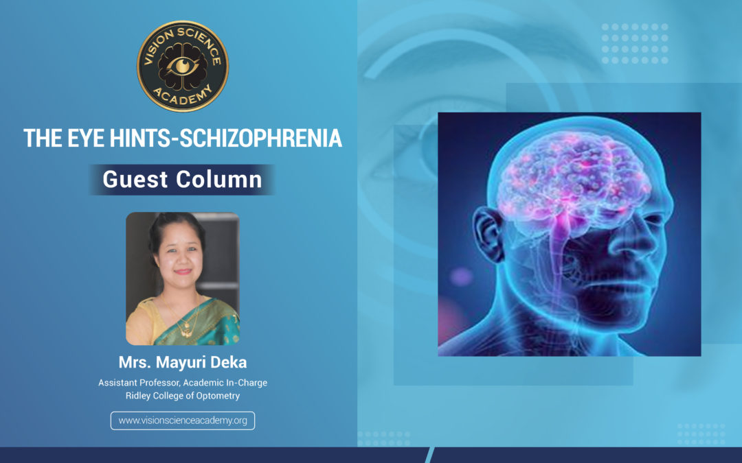
In the past decade, research has begun to point to a promising source of such signals: the eye. The thickness of a person’s retina or the retina’s response to light could provide early signs of schizophrenia. “The retina is essentially a proxy for what’s happening in the brain,” says Steven Silverstein, a clinical psychiatrist at Rutgers University in Piscataway, New Jersey. (1)
Visual processing impairments are well established in Schizophrenia, including abnormalities in contrast sensitivity. There has been little work on colour processing to date, but clinical reports indicate frequent descriptions of increased intensity, or alterations in colour perception. Visual distortions also have the highest predictive validity, among all basic symptoms for progressing to a psychotic disorder. Visual distortions in help seeking adolescents are associated with suicidal ideation, even after controlling for age, gender, depression, thought disorder, paranoia, and auditory distortions. (2) Finally, visual impairments contribute substantially to poorer real-world functioning in people with schizophrenia.
The potential to use the eye as a window to the brain goes beyond just schizophrenia. As understanding the links between eye and brain health deepens, evidence is building those changes in the eye can hint at the presence and progression of neurological disorders.
It was assumed that ‘Vision’ uses about half of the brain’s neural pathways. Anything that affects a person’s brain-whether a disease or a blow to the head-therefore has a strong chance of affecting their sight. Indeed, researchers have known for decades that certain neurological disorders can bring about changes in vision. There is evidence that, in people with schizophrenia, the small veins or venules, of the retina are wider and the retina is thinner. (3)
Perhaps, the most promising tool in the short term for identifying people at risk of developing schizophrenia is Electroretinogram (ERG), a simple and minimally invasive test that measures the retina’s electrical response to light. (4) That signal is captured using a small electrode that is attached to the cheek below the eye or placed under the upper or lower eyelid. Currently, non-invasive eye tracking systems using video cameras are available as well. Recent advances in the eye tracking cameras allow us to measure eye movements with high temporal and spatial resolution.

Figure 1: Eye movements reveal impaired reading in Schizophrenia
(Source – Images from Google Schizophrenia and Eyes, McGill University)
In the field of psychiatry, studies on eye movement characteristics in mental illness have been conducted since the early 1900s. Participants with schizophrenia were known to have characteristic eye movements during smooth pursuit, saccade control, and visual search. Studies have shown that in schizophrenia, eye movement measures such as scan path length, visual cognition such as perceptual organization, and social functioning measured by total work hours per week may have a hierarchical relationship, where eye movement characteristics lead to changes in cognition or social functioning.(5) Such findings are important because findings of hierarchical relationships between measures and social functioning have led to the development of new treatment and rehabilitation options.
In addition to acuity differences, other ocular changes related to schizophrenia, lack of good health care in people, and/or it’s treatment can affect performance on visual processing tasks. A study reported that a sample of schizophrenia patients had lens opacities, cataracts, and/or corneal pigmentation. Another condition more common in schizophrenia than the general population is strabismus. This can result in amblyopia, and depth perception difficulties, and people with schizophrenia have shown poor performance on visual tasks such as contour integration that resembles that of people with amblyopia as well as problems with depth perception. (1)
Conclusion
This article tries to highlight potential contributors to the visual processing abnormalities of schizophrenia and high-risk locations that are local to the retina or other structures of the eye. Therefore, to determine the retinal involvement, a more comprehensive understanding of the nature of the full range of visual processing and visual cognition impairments it needed.
References:
- Silverstein SM, Rosen R. Schizophrenia and the eye. Schizophrenia Research: Cognition. 2015 Jun 1;2(2):46-55.
- Woo M. Eyes hint at hidden mental-health conditions. Nature. 2019 Apr 10.
- Ahn J, Lee JY, Kim TW, Yoon EJ, Oh S, Kim YK, Kim JM, Woo SJ, Kim KW, Jeon B. Retinal thinning associates with nigral dopaminergic loss in de novo Parkinson disease. Neurology. 2018 Sep 11;91(11):e1003-12.
- Talman LS, Bisker ER, Sackel DJ, Long Jr DA, Galetta KM, Ratchford JN, Lile DJ, Farrell SK, Loguidice MJ, Remington G, Conger A. Longitudinal study of vision and retinal nerve fiber layer thickness in multiple sclerosis. Annals of neurology. 2010 Jun;67(6):749-60.
- Morita K, Miura K, Kasai K, Hashimoto R. Eye movement characteristics in schizophrenia: A recent update with clinical implications. Neuropsychopharmacology reports. 2020 Mar;40(1):2-9.
Author:-

Mrs. Mayuri Deka is currently working as an assistant professor and the academic in charge at the Ridley College of Optometry, Jorhat, Assam, India for the last 10 years. She has been actively involved in establishing optometry at the forefront of Assam’s eye care fraternity. Initially, the course of Optometry in Assam was not uniform or streamlined. Mayuri has contributed to streamlining the course and creating awareness amongst all about the noble profession. She has also delivered lectures/topics on Optometry like Sports Vision, Binocular Vision, etc. Additionally, she has also been a part of panel discussions in seminars/conferences and also spoken on Women empowerment on various occasions and platforms/webinars. Mrs. Mayuri is a leading member of the Assam Optometry Association.
Recent Comments