


Manvi Gupta, B. Optom., F. Optom (SCEH)
Adjunct Optometrist, Dr. Shroff’s Charity Eye Hospital, Delhi, India
Corneal Neuralgia is a serious but often undiagnosed condition. It typically presents after Laser-Assisted In Situ Keratomileusis (LASIK), which is used to correct myopia, hyperopia, and astigmatism by creating a flap and ablating the stroma to the desired extent. While cases of corneal neuralgia are not very often seen, they are usually the most troubling aspect in the life of the patient, despite a seemingly normal ocular examination. (1,2)
What is Corneal Neuralgia?
Corneal neuralgia is characterised by chronic ocular pain often arising from dysfunction or damage to the corneal nerves. (1,2) Unlike patients with dry eye, who suffer from ocular pain due to tear film instability, patients with corneal neuralgia suffer from persistent pain due to corneal nerve sensitisation. (4) Patients suffer from heightened responses even with non-painful stimuli and find it difficult to open their eyes in a closed, well-ventilated environment. (1)
Pathophysiology
During LASIK, a flap is created, which includes severing of corneal nerves and ablating the corneal stroma to the desired amount. In most of the patients, these corneal nerves regenerate over time, but in some rare cases, corneal sensitisation may occur, resulting in chronic pain. (1,2) More commonly seen in patients with dry eyes, autoimmune disorders, or psychological stress. (4,5)
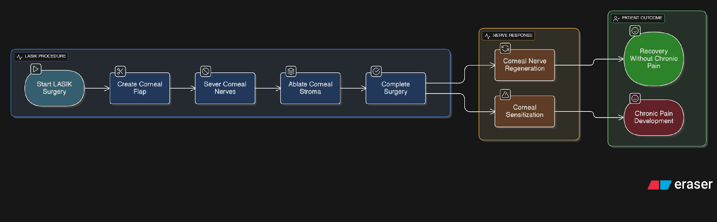

Figure 1: Steps Involved in LASIK Leading to Corneal Nerve Regeneration or Sensitisation
Symptoms
- Patients often experience persistent ocular pain. (1)
- Many report a foreign body sensation in the eye. (1)
- A common symptom is a burning sensation. (1,4)
- Individuals may also suffer from photophobia. (2)
- There is usually little to no relief from instilling lubricating agents. (1)
Diagnosis
Typically, diagnosis depends on clinical observation and the history of the patient. Normally, clinical findings may not correlate with the symptoms of the patient. (1) However, we can still perform certain diagnostic tests, like:
- Corneal Aesthesiometry: Aesthesiometer or cotton swab to assess corneal nerve response. (1)
- Topical Anaesthetic: After instilling anaesthetics, the pain persists. (2)
- Confocal Microscopy: To visualise corneal nerve damage. (3)
Management
Corneal Neuralgia is treated in a variety of ways, including:
- Topical treatments include topical cyclosporine, autologous serum eye drops, and artificial tears without preservatives. (1)
- Oral drugs: Neuropathic painkillers such as duloxetine, pregabalin, or gabapentin. (1)
- For the treatment of chronic pain, adjunct therapies include scleral lenses, punctal plugs, or Cognitive Behavioural Therapy (CBT). (1)
- New therapies include the investigation of botulinum toxin and transcranial magnetic stimulation. (1)
Conclusion
An under-diagnosed illness that can have a major influence on the quality of life of a patient is Corneal Neuralgia following LASIK. (1,2) Effective management requires early detection and a comprehensive approach to therapy. Understanding of clinicians can assist in closing the gap between symptoms and diagnosis, which will benefit those who are impacted. (1)
References
- Galor, A., Moein, H. R., Lee, C., Rodriguez, A., Felix, E. R., Sarantopoulos, C. D., & Levitt, R. C. (2015). Neuropathic ocular pain: An important yet underevaluated condition. Eye, 29(3), 301–312.
- Rosenthal, P., & Borsook, D. (2016). Ocular neuropathic pain. The British journal of ophthalmology, 100(1), 128–134.
- Midena, E., Cortese, M., Miotto, S., Gambato, C., Cavarzeran, F., & Ghirlando, A. (2009). Confocal microscopy of corneal sub-basal nerve plexus: a quantitative and qualitative analysis in healthy and pathologic eyes. Journal of refractive surgery (Thorofare, N.J. : 1995), 25(1 Suppl), S125–S130.
- Belmonte, C., Aracil, A., Acosta, M. C., Luna, C., & Gallar, J. (2004). Nerves and sensations from the eye surface. The ocular surface, 2(4), 248–253.
- Shaheen, B. S., Bakir, M., & Jain, S. (2014). Corneal nerves in health and disease. Survey of ophthalmology, 59(3), 263–285.