


Daksh Jain, B. Optom Student ,
Shubham Bhattacharya, Assistant Professor
Sumandeep Vidyapeeth (Deemed to be) University, Vadodara, India
Keywords
artificial intelligence, optometry, retinal imaging, clinical decision support, eye care.
Declaration of interest
This article is written for educational purposes and does not have any financial support or conflict of interest.
Introduction
There are 2.2 billion people with vision impairment right now. That is roughly one in every four humans on the planet. And here is the part that should bother you as a future eye care professional: a huge chunk of that is preventable.
If you are picturing a robot wearing a white coat and doing refractions, that is not what this is. Artificial Intelligence (AI) in eye care is more like having a ridiculously fast, never-tired assistant who can look at a retinal photo in 0.3 seconds and say, “Hey, you might want to look at this area more closely”.
How Artificial Intelligence Supports Clinical Decisions
AI is a type of computer system that can learn from examples. It can look at images such as fundus photographs and Optical Coherence Tomography scans. Once it has been trained on data, it can identify features that may be associated with disease. This helps doctors prioritise which patients need to be seen. If the image is not clear or if the patient has an unusual condition, the doctor may need to look at the results again. (1)
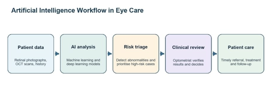

Figure 1. This image shows the workflow of Artificial Intelligence in eye care.
Where it is Useful?
One of the most important uses of AI is Diabetic Retinopathy screening. AI can detect fundus-image features such as microaneurysms, haemorrhage and exudates, helping identify patients who should be referred to a specialist. (2) It is also being used in Glaucoma care, where it can help detect changes in the optic nerve and retinal nerve fibre layer. (3) In addition, AI is being explored for predicting Myopia risk and for triaging images in tele-optometry. These applications may extend screening services to communities with limited access to specialists. Even so, the final decision to refer a patient should always rest with a doctor. (1,4)
Benefits, Limits and Responsibility
AI can make screening faster, more consistent and more accessible. However, its performance depends on good-quality images, representative training data and validation in the population where it will be used. Poor data may lead to missed disease or unnecessary referrals. Clinicians must also consider privacy, data security, and algorithmic bias. For this reason, AI should be treated as additional evidence rather than a final diagnosis. (1,4)
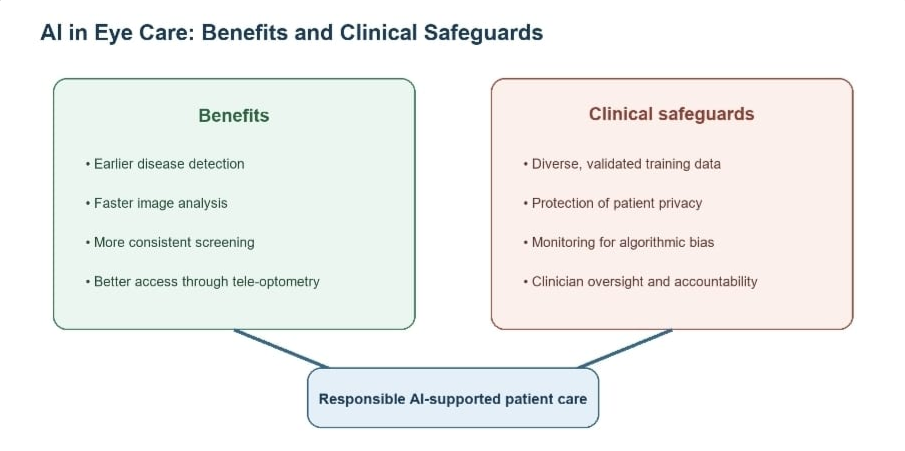

Figure 2. This image shows the benefits and challenges of AI in vision sciences.
Why this Matters to Optometrists?
You are graduating into a profession that is already changing. AI will not replace you, but Optometrists who use AI effectively will replace those who do not. Here is where you will actually encounter it.
Therefore, where will you actually meet AI in practice? Everywhere but never in the driver’s seat. In refraction, it will suggest a starting Rx from autorefractor data and history, but it cannot know your patient is a 55-year-old accountant who will hate that “perfect” -0.50D cylinder at 40cm.
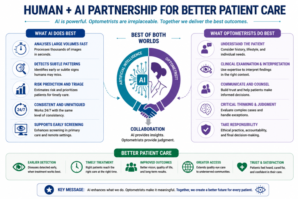

Figure 3. This image shows the complementary roles of AI and Optometrists in delivering patient care.
Conclusion
AI has the potential to improve detection and access to eye care especially when clinical services are busy. The future of Optometry is not about doctors versus technology; it is about doctors using technology responsibly.
References
- Ting DSW, Pasquale LR, Peng L et al. Artificial intelligence and Deep learning in ophthalmology. Br J Ophthalmol. 2019;103(2):167-175. Doi:10.1136/bjophthalmol-2018-313173.
- Gulshan V, Peng L, Coram M et al. Validation of a deep learning algorithm for detection of diabetic retinopathy in retinal fundus photographs. JAMA. 2016;316(22):2402-2410. Doi:10.1001/jama.2016.17216.
- Li Z, He Y, Keel S, et al. Efficacy of a deep learning system for detecting optic neuropathy based on color fundus photographs. Ophthalmology. 2018;125(8):1199-1206. Doi:10.1016/j.ophtha.2018.01.023.
- Kapoor R, Walters SP, Al-Aswad LA. The current state of artificial intelligence in ophthalmology. Surv Ophthalmol. 2019;64(2):233-240. Doi:10.1016/j.survophthal.2018.07.002.
About the Authors


Daksh Jain
B. Optom Student ,
Sumandeep Vidyapeeth (Deemed to be) University, Vadodara, India

Shubham Bhattacharya
Assistant Professor ,