

Haziel rynjah
Assistant Professor, T
The Assam Royal Global University, Guwahati, India

Deepjyoti Baro
Clinical Optometrist,
Sri Sankaradeva Nethralaya, Guwahati, India
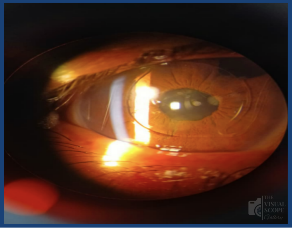
Figure: Slit-lamp photograph demonstrating an intraocular lens (IOL) with a peripheral iridectomy in a pseudophakic eye during postoperative follow-up.
Image Summary
The patient presented for routine ocular follow-up with a history of iridectomy and intraocular lens implantation in 2025. Visual acuity in the affected eye was 6/12. A slit-lamp examination revealed a pseudophakic eye with a visible intraocular lens and a superior peripheral iridectomy. The IOL appeared to be well placed, and there was no apparent significant anterior segment abnormality in the attached photograph. The case is representative of the characteristic appearance of the anterior segment after iridectomy with IOL implantation. The postoperative status should be followed up by assessment of visual acuity, position of IOL, corneal status, anterior chamber, iridectomy site, intraocular pressure, and posterior segment.
Recent Comments