
Tanvi Mahendra Gaonkar, M. Optom
Assistant Professor, ITM Vocational University, Vadodara, India
Migraine is a complicated neurovascular disease that involves activation of the trigeminovascular system, changes in blood flow, and varied brain activity. (1) Recent studies have shown that Migraine is not just a brain disorder but also a condition associated with dysfunction in the microcirculation and systemic vascular changes. (2) The blood supply in the retina and autoregulatory systems are identical to those in the brain, as both were developed from the same embryonic tissue. However, unlike cerebral vessels, retinal vessels are easily and non-invasively visible. (1)
With the development of Optical Coherence Tomography (OCT) and Optical Coherence Tomography Angiography (OCTA), clinicians can now examine retinal structure and microvasculature in detail. Studies increasingly show measurable retinal neural and vascular changes in people with Migraine. (3) These findings position the retina as a valuable window into Migraine-related neurovascular health.
Migraine and Vascular Dysregulation
Migraines involve a complex relationship between the neural and vascular systems. Cortical spreading depression is a major mechanism where a wave of changed electrical activity moves across the brain cortex. This leads to temporary variations in cerebral blood flow. (4) Flashing lights and zigzag patterns are examples of visual aura symptoms. Another important pathway is trigeminovascular activation. This process releases vasoactive substances that affect blood vessel tone and contribute to pain and vascular instability. These support the idea that migraine is associated with broader vascular dysregulation, which may also affect the retinal circulation.
Retina as a Reflection of Brain Microcirculation
The retina is considered an extension of the central nervous system. It originates from the neural tube. It shares structural and functional similarities with the brain. Retinal vessels and cerebral vessels both rely on tight autoregulation to maintain consistent blood flow. (5) Changes seen in the retina may show similar microvascular processes occurring in the brain. (1)
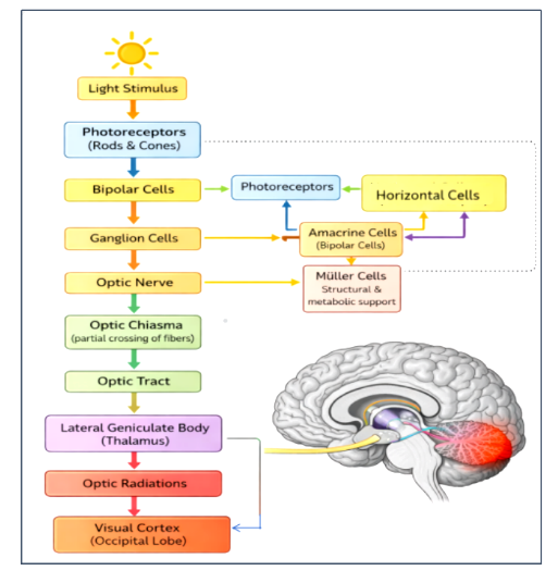
Figure 1: This image shows retina–brain connection illustrating retina as a window to neurodegeneration.
Image Courtesy: Created by the Author
Optical Coherence Tomography Angiography (OCTA) Findings
A systematic review and meta-analysis by a study that included 775 eyes and findings showed reduced superficial and deep macular vessel density, decreased peripapillary vessel density, and enlargement of the Foveal Avascular Zone (FAZ) in Migraine patients. These signs were more severe in patients with Migraine with aura. (1) Changes in retinal blood flow are indicated by reduced vessel density. (5)
However, an increase in the FAZ shows a decrease in capillary perfusion in the macular region. (4) Another study investigated retinal neural tissue thickness and vascular calibres in individuals with Migraine. Although large differences were not consistently observed, subtle variations in retinal nerve fibre layer thickness and vascular measurements were noted. (2) Some Migraine subgroups also showed retinal neural layer thinning. (7) These results can be a sign of periodic episodes of vascular imbalance over time.
Conclusion
In conclusion, Migraine is recognised as a disorder of neurovascular instability. Reduced vessel density, enlargement of the FAZ, and subtle structural alterations highlight the close relationship between cerebral and retinal circulation. The retina truly serves as a window to understanding Migraine.
References
- Pang, Y., Cao, T., Zhang, Q., Hu, H., Wang, Z., Nie, J., … & Zhang, X. (2023). Retinal microvasculature features in patients with migraine: a systematic review and meta-analysis. Frontiers in neurology, 14, 1187559.
- Ristioja, S., Leiviskä, I. L., Saarela, V. O., & Liinamaa, M. J. (2024). Retinal neural tissue and vascular calibres in migraine: the Northern Finland Birth Cohort Eye Study. Acta Ophthalmologica, 102(5), 600-609.
- Chaliha, D. R., Vaccarezza, M., Charng, J., Chen, F. K., Lim, A., Drummond, P., … & Mamo, J. C. (2024). Using optical coherence tomography and optical coherence tomography angiography to delineate neurovascular homeostasis in migraine: a review. Frontiers in neuroscience, 18, 1376282.
- Tukur, H. N., Uwishema, O., Sheikhah, D., & Akbay, H. (2025). Neuro-ophthalmology and migraine: visual aura and its neural basis. International Journal of Emergency Medicine, 18(1), 148.
- Wang, L., Shah, S., Llaneras, C. N., & Goldhardt, R. (2024). Insight into the brain: application of the retinal microvasculature as a biomarker for cerebrovascular diseases through optical coherence tomography angiography. Current ophthalmology reports, 12(1), 1-11.
- Sijilmassi, O. (2026). The Retina as a Proxy for Brain Neurodegeneration: A Narrative Review on OCT-Based Retinal Imaging in the Early Detection of Alzheimer’s and Parkinson’s Disease. Journal of Imaging, 12(3), 104.
- Conway, M. L., & Ctori, I. (2023). Retinal and choroidal alterations in migraine patients compared to normal healthy controls. Journal of Behavioral and Brain Science, 13(10), 185-197.
About the Author

Tanvi Mahendra Gaonkar
Assistant Professor
Recent Comments