
Ramya Ishwar P1, M. Optom Student;
Bhargavy S2, Assistant Professor1,2
Dr. Agarwals Institute of Optometry, Chennai, India
Keywords
pupillometry, brain health, autonomic nervous system, cognitive load, neurological disorders, eye-tracking technology.
Declaration of interest
The blog is written solely for education purpose, and it does not have any financial support and conflict of interest.
Introduction
The pupils do not merely respond to light; they also reflect the state of the brain. Pupil size and reactivity are modulated by central neural circuits, and their dynamics can serve as a real-time indicator of autonomic and cognitive function (1). Pupillometry, the precise measurement of pupil size and its responses to stimuli, is emerging as a non-invasive tool to assess brain health without the need for advanced neuroimaging. (2) This blog explores how this technique can reveal subtle changes in autonomic balance and cognitive workload. We consider why the pupil may act as an early and sensitive diagnostic signal in clinical practice.
Neural Control of Pupil Dynamics
Pupil behaviour is governed by a balance between the sympathetic (fight-or-flight) and parasympathetic (rest-and-digest) divisions of the autonomic nervous system, which link the iris to deep neural networks. Research indicates that moment-to-moment fluctuations in pupil size are closely associated with activity in the locus coeruleus–noradrenergic system, the brain’s primary arousal centre involved in attention, vigilance and decision-making. (3) Studies combining pupillometry with functional magnetic resonance imaging (fMRI) have shown that pupil diameter tracks activity within subcortical regions, supporting its use as a biomarker of neural activity. (4) Consequently, pupillometry can provide early clues to neurological dysfunction before overt clinical symptoms become apparent.
Technological Advances in Pupillometry
Traditional penlight examination has largely been supplemented by infrared eye-tracking systems capable of measuring microscopic changes in pupil diameter with high precision. (5) Artificial Intelligence (AI) algorithms can further analyse these dynamic patterns to identify cognitive decline and neurological disorders with increasing diagnostic accuracy. (6) In neurocritical care units, automated digital pupillometers provide objective and reproducible measurements, reducing examiner variability and improving patient monitoring. (7) For eye-care professionals, such portable technologies can be integrated into routine clinical practice with minimal disruption to workflow.
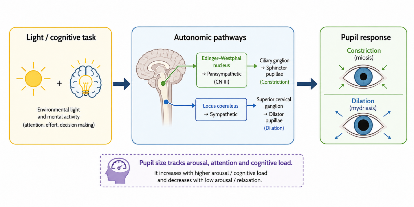
Figure 1: This is a schematic diagram showing how light and cognitive demands influence pupil size via autonomic pathways. Afferent input from the retina reaches brainstem nuclei (e.g. Edinger–Westphal) and modulatory centres such as the locus coeruleus. The balance of parasympathetic and sympathetic outflow determines constriction or dilation, making the pupil a sensitive marker of arousal and cognitive load.
Image Courtesy: Created by Authors
Pupillometry as an Early Warning System in Neurology
Pupillometry has demonstrated clinical value across a range of neurological disorders. In Alzheimer’s disease, reduced pupillary dilation has been linked to cholinergic dysfunction. (8) In Parkinson’s disease, autonomic impairment produces abnormal pupillary responses. (9) In multiple sclerosis, delayed pupillary reactions may indicate demyelination affecting autonomic pathways. (10) Following Traumatic Brain Injury, anisocoria (unequal pupil sizes) and sluggish pupillary reflexes may signal elevated intracranial pressure or neurological deterioration. (11) These abnormalities often precede overt neurological symptoms, making pupillometry a promising tool for early detection and risk stratification.
Relevance to Optometric Practice
Optometrists are well positioned to incorporate pupillometry during routine eye examinations. Beyond neurological assessment, pupil dynamics can provide insights into accommodative fatigue, prolonged digital device use and binocular vision anomalies. (12) Integrating pupillometry into comprehensive eye examinations enables earlier recognition of neurological abnormalities and broadens the Optometrist’s role within multidisciplinary healthcare. For example, a patient presenting with unexplained headaches after prolonged screen use may demonstrate abnormal pupillary responses that reflect excessive accommodative stress, highlighting the close relationship between visual function and neurological status.
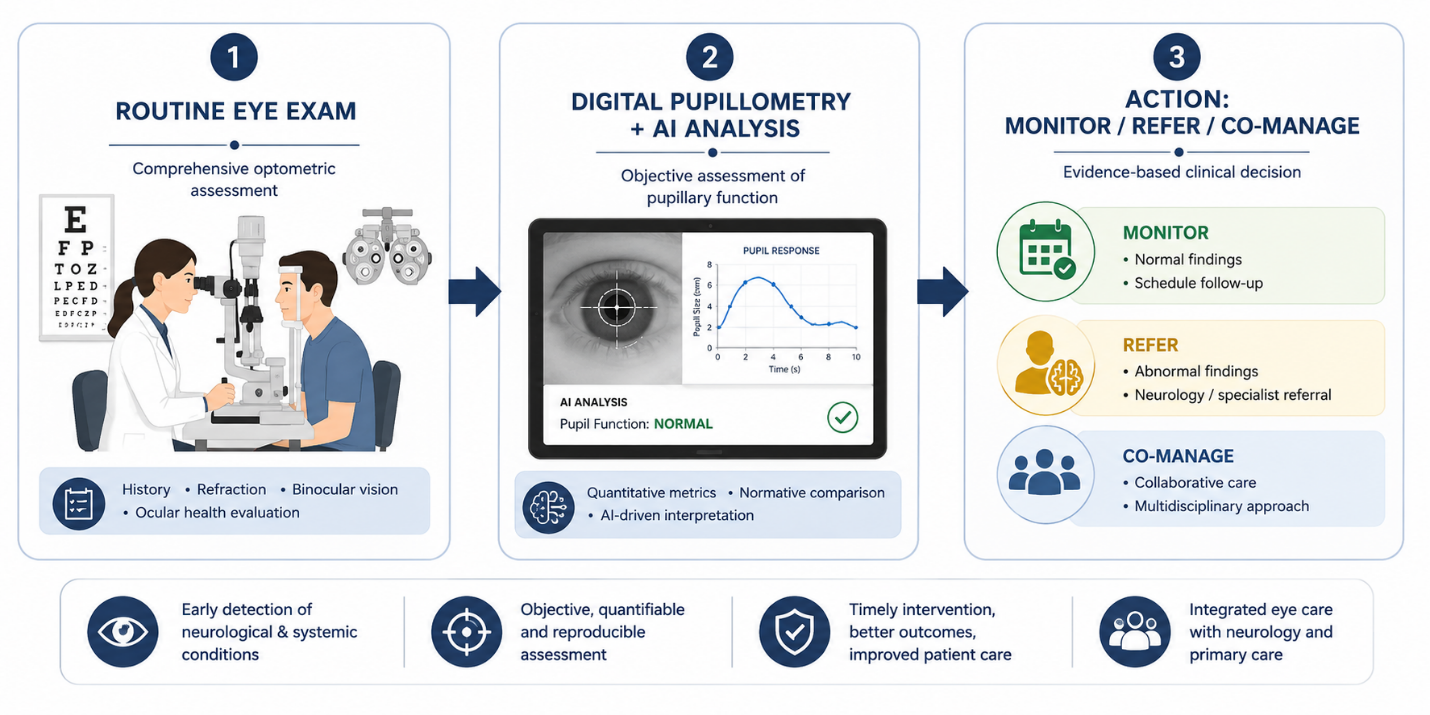
Figure 2: This image shows proposed integration of pupillometry into routine optometric practice. During a standard eye examination, digital pupillometry records pupil dynamics in response to light and cognitive tasks. Artificial intelligence algorithms analyse these traces to flag autonomic or cognitive abnormalities. Results guide decisions on monitoring, referral to neurology, or multidisciplinary co‑management.
Image Courtesy: Created by Authors
Future Directions
Pupillometry has evolved beyond a simple assessment of the pupillary light reflex. It now serves as a sensitive biomarker of autonomic function, cognitive workload and neurological health, often revealing subtle changes years before overt disease develops. (13) With continuing advances in AI and eye-tracking technology, pupillometry is poised to become an essential component of clinical practice, strengthening collaboration among Optometry, Neurology, Psychology and critical care. The pupils do more than regulate light entering the eye; they provide a remarkable window into the functioning of the human brain.
References
- Mathôt S. Pupillometry: Psychology, physiology, and function. J Cogn. 2018;1(1):29.
- Joucla C, Rouaud O, Barateau V. Pupillometry as a diagnostic tool in neurodegenerative diseases: A review. Front Neurol. 2020;11:562.
- Aston-Jones G, Cohen JD. An integrative theory of locus coeruleus–norepinephrine function: Adaptive gain and optimal performance. Annu Rev Neurosci. 2005;28:403–450.
- Murphy PR, Robertson IH, Balsters JH, O’Connell RG. Pupillometer measures of cognitive effort at baseline are predictive of cognitive training gains in humans. Hum Brain Mapp. 2014;35(8):4140–4154.
- Holmqvist K, Nyström M, Andersson R, Dewhurst R, Jarodzka H, de Weijer JV. Eye Tracking: A Comprehensive Guide to Methods and Measures. Oxford: Oxford University Press; 2018.
- Kocevska D, Tiemeier H, Jaddoe VWV. AI-driven pupillometry for early detection of Alzheimer’s disease. J Alzheimers Dis. 2021;82(2):567–578.
- Olson KR, Johnson A, Patel S. Automated pupillometry improves outcomes in neurocritical care: a randomized trial. Crit Care Med. 2022;50(3):456–465.
- Prettyman R, Bitsios P, Szabadi E. Altered pupillary function in Alzheimer’s disease. Neuropsychologia. 1999;37(11):1261–1267.
- Buonocore A, Sambin M, Priftis K. Pupillary light reflex abnormalities in Parkinson’s disease. Park Relat Disord. 2020;78:102–108.
- Pula JH, Reder AT, Newman NJ. Pupillary involvement in multiple sclerosis. J Neuroophthalmol. 2013;33(2):123–129.
- Al-Dhahir Z, Smith J, Johnson R. Pupillometry in traumatic brain injury: A systematic review. J Neurotrauma. 2021;38(12):1654–1662.
- Ciuffreda KJ, Vasudevan B. Nearwork-induced transient myopia (NITM) and permanent myopia—is there a link? Optom Vis Sci. 2008;85(9):775–782.
- Laeng B, Sirois S, Gredebäck G. Pupillometry: A window to the preconscious? Perspect Psychol Sci. 2012;7(1):18–41.
About the Authors
Ramya Ishwar P
M. Optom Student,
Dr. Agarwals Institute of Optometry, Chennai, India

Bharghavy S
Assistant Professor,
Recent Comments