
Alaa Mohammed Abdallh Mohammed, B. Sc. Optometry Student
Faculty of Optical Sciences, Neelain University, Sudan
Keywords
alzheimer’s disease, retinal imaging, optical coherence tomography, artificial intelligence, neurodegeneration
Declaration of Interest
The blog is written solely for educational purposes and has no financial support or conflict of interest
What if a person could notice subtle changes in vision years before memory problems became obvious? Imagine an ordinary eye examination revealing clues that might prompt further neurological assessment. This possibility is driving growing interest in the retina as a potential window into brain health.
Alzheimer’s and the Case for Early Detection
Alzheimer’s Disease (AD) is the most common cause of dementia worldwide and is characterised by progressive memory loss and cognitive decline. By the time symptoms become noticeable, significant neuronal damage may have already occurred. Therefore, researchers are focusing on identifying early biomarkers that can detect individuals at risk before clinical symptoms appear.
Early detection is therefore not only a scientific priority but also a clinical one, as it may open a window for timely intervention and better long-term outcomes.
The Retina: A Window to the Brain
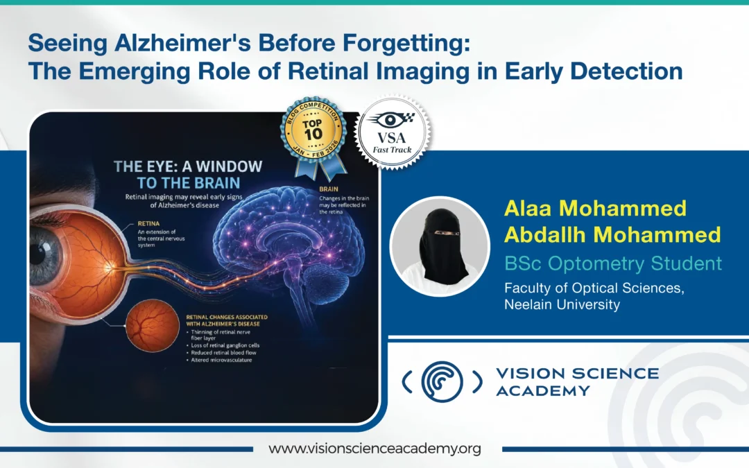
The retina is an extension of the central nervous system and shares important developmental and biological features with the brain. Unlike the brain, however, the retina can be examined directly and non-invasively. This makes retinal imaging an attractive approach for investigating whether ocular changes can reflect early neurodegenerative processes. (1)
Early Signs in the Retina
Research has reported retinal abnormalities in people with Alzheimer’s disease and mild cognitive impairment. These include thinning of the retinal nerve fibre layer, changes involving retinal ganglion cells, and alterations in retinal microvasculature. Such findings have raised interest in whether retinal measurements could provide clues about neurological health, although their diagnostic significance is still being investigated. (2,3)
Imaging Technology: OCT and OCTA
Optical Coherence Tomography (OCT) provides detailed cross-sectional images of retinal layers and has been investigated for detecting subtle structural changes associated with Alzheimer’s disease. Optical Coherence Tomography Angiography (OCTA) can also visualise retinal microvasculature without dye injection, providing complementary information about vascular changes. (4)
Artificial Intelligence and Pattern Recognition
Artificial Intelligence (AI) may further expand the potential of retinal screening. Machine-learning methods can analyse large collections of retinal images and identify patterns that may be difficult to recognise clinically. Emerging research suggests that AI-assisted retinal analysis could eventually support risk assessment and help identify people who may benefit from further neurological evaluation. (3)
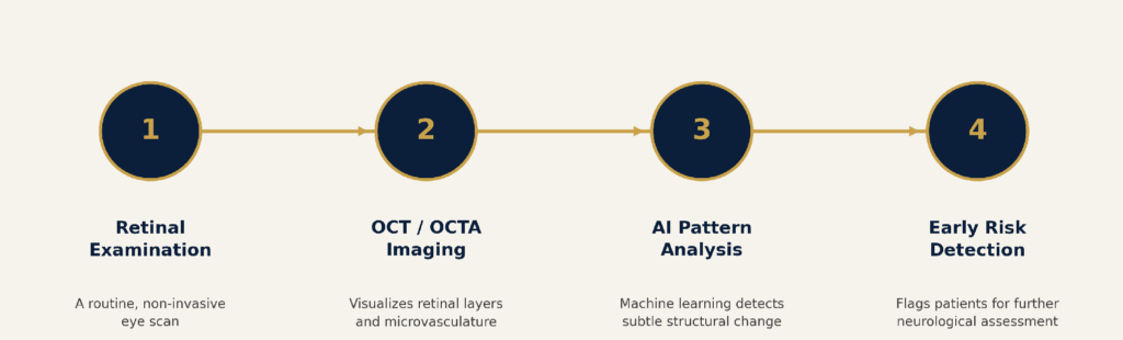
Figure 2: This image shows a proposed pathway from retinal examination and OCT/OCTA imaging to AI-assisted pattern analysis and further assessment.
Image Courtesy: Author-created visual.
“Machine learning algorithms can analyse large numbers of retinal images and identify complex patterns that may not be visible to clinicians.”
Current Limitations
However, retinal imaging is not yet a standalone diagnostic test for Alzheimer’s disease. Retinal changes may also occur due to normal ageing or other ocular and neurological conditions. Further large-scale, long-term studies are required to validate retinal biomarkers and establish reliable screening approaches.
Standardisation of imaging protocols and agreement on which retinal parameters are most meaningful will also be essential before such approaches can be adopted in routine clinical practice.
Looking Ahead
The future of Alzheimer’s detection may involve a combination of retinal imaging, AI, and neuroscience. A simple eye examination may one day provide an opportunity to identify neurodegenerative changes earlier, allowing timely intervention and improved patient care.
For now, retinal imaging should be viewed as a promising research tool rather than a routine screening method. Continued collaboration between Ophthalmology, Neurology, and Data Science will be key to translating these early findings into meaningful clinical benefit. (3)
For eye-care professionals, this emerging field highlights the importance of careful retinal assessment and accurate documentation. It also illustrates how Optometry may contribute to interdisciplinary care by recognising findings that warrant referral, while avoiding the assumption that a retinal abnormality alone confirms Alzheimer’s disease.
As evidence develops, collaboration between Optometrists, Ophthalmologists, Neurologists, and Researchers will remain important.
References
- London A, Benhar I, Schwartz M. The retina as a window to the brain—from eye research to CNS disorders. Nature Reviews Neurology. 2013;9(1):44–53.
- den Haan J, Verbraak FD, Visser PJ, Bouwman FH. Retinal thickness in Alzheimer’s disease: A systematic review and meta-analysis. Alzheimer’s & Dementia. 2017;6:162–170.
- Snyder PJ, Alber J, Alt C, et al. Retinal imaging in Alzheimer’s and neurodegenerative diseases. Alzheimer’s & Dementia. 2021;17(1):103–111.
- Chan VTT, Sun Z, Tang S, et al. Spectral-domain optical coherence tomography measurements in Alzheimer’s disease. Ophthalmology. 2019;126(4):497–510.
About the Author

Alaa Mohammed Abdallh Mohammed
BSc Optometry Student,
Recent Comments