
Achint Kaur, M. Optom
Consultant Optometrist and Educator, Silver Line Laboratories, New Delhi, India
When your patient removes the lenses in the morning and reads 6/6. Does that mean that the lenses you dispensed have worked properly?
Visual acuity is the ability of the eyes to see the details at various distances. It is the ability of the eyes to read a text, recognise a face, and identify objects both near and far, but visual acuity will not tell how the cornea has changed its shape to produce that vision. Corneal topography shows us the proper mapping of the cornea and helps to visualise the curvature changes and distribution of the Ortho-K treatment effect.
What Exactly Does a Corneal Topography Map Show?
In Myopic correction, the central cornea undergoes controlled flattening, accompanied by reversible redistribution of epithelial thickness. Surrounding this treatment zone, the paracentral cornea becomes relatively steeper, creating an annular pattern. The amount of this peripheral steeping is closely associated with the degree of central flattening required to correct a patient’s Myopia. (1)
A corneal topography map is essentially a 3D-like representation of the cornea’s surface, showing how its curvature and shape vary from the centre toward the periphery.
- Baseline Corneal Shape: Central corneal curvature, amount and orientation of corneal astigmatism, eccentricity, and any other irregularities that affect the lens fitting.
- Treatment Zone: Central corneal flattening corresponding to the optical/pupil zone.
- Return Zone Depth: Paracentral area of steeping surrounding the flattened zone. Mainly associated with the redistribution of corneal epithelial cellular fluid during myopia treatment.
- Treatment Zone Diameter: Very essential finding if the treatment zone is too small or too large may have implications for visual quality.
The Ortho-K Treatment Pattern: The “Fingerprint” of Corneal Reshaping
Ortho-K lenses are used for two main purposes: Myopia correction and Myopia control. (2,3) Ortho-K “fingerprint” refers to distinctive corneal topography pattern post lens wear. This pattern represents the individual corneal response to the Ortho-K treatment and reflects the interaction of several factors, including:
- Lens geometry
- Tear-film forces and fluid dynamics
- Pre-existing corneal shape
- Corneal epithelial cellular fluid redistribution
- Lens centration and movement
Ortho-K lenses produce a recognisable pattern on tangential or curvature maps, showing how the cornea has reacted overnight with the lens wear.
Typically, a pattern should be a bull’s eye pattern, where the central cornea is flattened, and peripheral cornea is steepened. But this pattern is more than just a visual signature. Each zone tells us something about how the Ortho-K lens has interacted with the cornea, while the position and shape of the treatment zone can provide valuable information about treatment centration and the overall reshaping response.
Various zones of Ortho-K pattern:
- Central zone: Treatment zone
- Mid peripheral zone: Return zone depth
- Landing zone: to maintain the reverse zone/Mid-periphery zone
- Edges: for adequate movement and centration (2,3)
Optical Coherence Tomography (OCT) based epithelial thickness mapping helps explain the structural basis of many of the changes observed in corneal topography. (4,7)
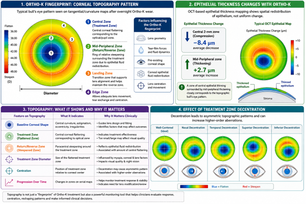
Figure 1: This image shows the original infographic created for this article using AI-generated medical illustration.
Image Courtesy: Created by the Author
In Ortho-K treatment, the epithelium does not uniformly become thicker or thinner; it undergoes spatial redistribution. At the centre of the cornea, epithelial thinning is typically observed. In a study involving 60 myopic eyes reported an average decrease of approximately 8.4 μm in epithelial thickness within the central 2-mm zone, accompanied by an increase of around 2.7 μm in the mid-peripheral epithelium. Notably, epithelial thickness mapping demonstrated a zone of central epithelial compression encircled by a ring of mid-peripheral thickening. This distribution showed a close relationship with the characteristic tangential topographic pattern produced by Ortho-K treatment. (4) Not all treatment zones are equal. The diameter and the shape of the treatment zone solely depend on corneal characteristics and lens design. The visual experience of the patient is also dependent on how much the pupil lies between the treatment zone, therefore, understanding the treatment zone plays a vital role. A 2023 study involving 106 eyes found that the treatment-zone dimensions were associated with the amount of myopia, corneal characteristics, and contact-lens parameters. The study also found that a larger back optic zone diameter was associated with larger treatment zones. (5) Topography also helps to find out whether the lens is well centred, shifted nasally, shifted temporally, and shifted superiorly/inferiorly. A well-centred treatment zone generally produces a more symmetrical pattern of central flattening surrounded by mid-peripheral steepening. In contrast, when the treatment zone is displaced nasally, temporally, superiorly, or inferiorly, the resulting topographic pattern may become asymmetric, with the areas of flattening and surrounding steepening shifted away from the corneal centre. (8,10)
The authors analysed topography from 26 myopic Ortho-K wearers and determined treatment centres from tangential maps using ellipse fitting. They found that changes in lens/treatment position were associated with decentration and that increasing decentration was associated with increasing higher-order aberrations. (6)
Topography is not just a “fingerprint” of Ortho-K treatment but also an important monitoring tool. Serial topographic maps help clinicians assess treatment progression, centration, refractive stability, and corneal changes, while also indicating when lens modification or further evaluation may be required.
Conclusion
Therefore, visual acuity just tells us about how well a patient sees; corneal topography tells us why. In Ortho-K, achieving good visual acuity does not necessarily indicate that the treatment zone is well centred, appropriately sized, or symmetrically formed. Corneal topography provides a more detailed assessment of the corneal changes induced by the lens, allowing practitioners to evaluate treatment response, centration, reshaping patterns, and any potential irregularities.
References
- Faria-Ribeiro M, Navarro Belsue R, López-Gil N, González-Méijome JM. Morphology, topography, and optics of the orthokeratology cornea. J Biomed Opt. 2016;21(7):075011. doi:10.1117/1.JBO.21.7.075011.
- Swarbrick HA. Orthokeratology (corneal refractive therapy): what is it and how does it work? Eye Contact Lens. 2004;30(4):181-5. doi:10.1097/01.icl.0000140221.41806.6e.
- Swarbrick HA, Wong G, O’Leary DJ. Corneal response to orthokeratology. Optom Vis Sci. 1998;75(11):791-9. doi:10.1097/00006324-199811000-00019.
- Kim WK, Kim BJ, Ryu I-H, Kim JK, Kim SW. Corneal epithelial and stromal thickness changes in myopic orthokeratology and their relationship with refractive change. PLoS One. 2018;13(9):e0203652. doi:10.1371/journal.pone.0203652.
- Gruhl J, Widmer F, Nagl A, Bandlitz S. Factors influencing treatment zone size in orthokeratology. Cont Lens Anterior Eye. 2023;46(4):101848. doi:10.1016/j.clae.2023.101848.
- Eltantawy E, Field E, Bui C, Kang M. Orthokeratology decentration: topography alignment, tear profile symmetry and induced HOAs. Ophthalmic Physiol Opt. 2026;46(2):366-78. doi:10.1007/s44402-026-00046-y.
- Alharbi A, Swarbrick HA. The effects of overnight orthokeratology lens wear on corneal thickness. Invest Ophthalmol Vis Sci. 2003;44(6):2518-2523. doi:10.1167/iovs.02-0680.
- Maseedupally VK, Gifford P, Lum E, Naidu R, Sidawi D, Wang B, et al. Treatment zone decentration during orthokeratology on eyes with corneal toricity. Optom Vis Sci. 2016;93(6):609-17. doi:10.1097/OPX.0000000000000896.
- Hiraoka T, Mihashi T, Okamoto C, Okamoto F, Hirohara Y, Oshika T. Influence of induced decentred orthokeratology lens on ocular higher-order wavefront aberrations and contrast sensitivity function. J Cataract Refract Surg. 2009;35(11):1918-26. doi:10.1016/j.jcrs.2009.06.018.
- Zhang Y, et al. The treatment zone decentration and corneal refractive profile changes in children undergoing orthokeratology treatment. BMC Ophthalmol. 2022;22:146. doi:10.1186/s12886-022-02310-4
About the Author

Achint Kaur
Consultant Optometrist and Educator,
Recent Comments