

Gomathi Suresh, B. Optom
Sr. Assistant Professor and Multimedia Manager, Dr. Agarwals Institute of Optometry, Chennai, India
Meibomian Gland Dysfunction (MGD) is a leading cause of evaporative dry eye disease and a significant contributor to ocular discomfort worldwide. Its prevalence increases with age and prolonged use of digital devices, yet it often remains underdiagnosed due to variability in clinical assessment. Symptomatic irritation of the eyelid and globes as well as decreased visual acuity has been reported. (1) Early and accurate grading of MGD is crucial for timely management and prevention of chronic dry eye complications.
Challenges in Conventional MGD Assessment
Traditional evaluation of MGD relies on slit-lamp examination of lid margins, assessment of meibomian gland orifices, impressibility, and secretion quality. These assessments are largely subjective and dependent on clinician experience, leading to inter-observer variability. (2) In busy clinics and community screening settings, detailed lid margin assessment may be overlooked, resulting in underdiagnosis.
Role of Artificial Intelligence in MGD Grading
Artificial Intelligence (AI), particularly machine learning and deep learning algorithms, offers a novel approach to standardising MGD assessment. AI systems trained on high-quality images of upper eyelid meibomian gland orifices can automatically detect morphological changes such as orifice plugging, pouting, telangiectasia, and lid margin irregularities. (3)
By converting visual patterns into objective scores, AI enables automated grading of MGD severity, reducing subjectivity and improving consistency across examiners and clinical settings.
AI-Based Workflow for MGD Screening
An AI-driven MGD grading system follows a simplified workflow suitable for clinics and screening programs. (Figure 1)
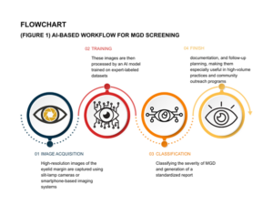
Figure 1: This image shows the flowchart for MGD Screening
Image Courtesy: Created by the Author
Improving Clinical Efficiency and Patient Care
AI-driven MGD grading improves clinical efficiency by reducing examination time and assisting clinicians in decision-making. (4) Objective grading supports treatment selection, monitoring of disease progression, and patient education. Visual AI outputs also help patients better understand their condition, improving compliance with lid hygiene and treatment regimens.
Limitations and Ethical Considerations
Despite its potential, AI-based MGD grading faces limitations such as dependence on image quality, variability in lighting and eyelid eversion techniques, and limited representation of diverse populations in training datasets. (5-6) Ethical considerations including data privacy, informed consent, and algorithm transparency are essential to ensure responsible implementation.
Conclusion
AI-driven grading of Meibomian Gland Dysfunction represents a new frontier in dry eye management. By offering objective, consistent, and scalable assessment, AI has the potential to transform how MGD is diagnosed and monitored. When integrated thoughtfully with clinical expertise and ethical safeguards, AI can enhance early detection, optimise treatment strategies, and ultimately improve the quality of life for patients with dry eye disease
References
- Chhadva P, Goldhardt R, Galor A. Meibomian Gland Disease: The Role of Gland Dysfunction in Dry Eye Disease. Ophthalmology. 2017 Nov;124(11S):S20-S26. doi: 10.1016/j.ophtha.2017.05.031. PMID: 29055358; PMCID: PMC5685175.
2. Sullivan BD, Smith GT, Gupta A, Harman F, Ansari E. Impact of Clinician Subjectivity on the Assessment of Dry Eye Disease Prevalence in a UK Public Health Care Patient Population. Clin Ophthalmol. 2024 Mar 8;18:743-753. doi: 10.2147/OPTH.S452149. PMID: 38476359; PMCID: PMC10929644.
3. Q. Dai et al., “A Novel Meibomian Gland Morphology Analytic System Based on a Convolutional Neural Network,” in IEEE Access, vol. 9, pp. 23083-23094, 2021, doi: 10.1109/ACCESS.2021.3056234.
4. Zhang YY, Zhao H, Lin JY, Wu SN, Liu XW, Zhang HD, Shao Y, Yang WF. Artificial Intelligence to Detect Meibomian Gland Dysfunction From in-vivo Laser Confocal Microscopy. Front Med (Lausanne). 2021 Nov 25;8:774344. doi: 10.3389/fmed.2021.774344. PMID: 34901091; PMCID: PMC8655877.
5. Li L, Xiao K, Lai T, Lai K, Lin J, Ge Z, Liang L, Huang H, Zhang X, Liu L, Wang Y, Shang X, He M, Xue Y, Zhu Z. Development and multicenter validation of an AI driven model for quantitative meibomian gland evaluation. NPJ Digit Med. 2025 Jul 4;8(1):403. doi: 10.1038/s41746-025-01753-5. PMID: 40615666; PMCID: PMC12227535.
6. Schraml D, Notni G. Synthetic Training Data in AI-Driven Quality Inspection: The Significance of Camera, Lighting, and Noise Parameters. Sensors (Basel). 2024 Jan 19;24(2):649. doi: 10.3390/s24020649. PMID: 38276341; PMCID: PMC10820774.
About the Author

Gomathi Suresh
Sr. Assistant Professor and Multimedia Manager,
Recent Comments