
Mohammad Akif Akram, B. Optom
Adjunct Optometrist, Dr. Shroff’s Charity Eye Hospital, New Delhi, India
Imagine restoring the sight of a blind person using their own tooth. As extraordinary as it sounds, Osteo-Odonto-Keratoprosthesis (OOKP), commonly known as “tooth-in-eye” surgery, is a life-changing procedure for patients with severe corneal blindness when all other treatments have failed. In OOKP, surgeons use the canine tooth of the patient and surrounding jawbone to support a tiny transparent optical lens. This tooth-bone structure is then implanted into the eye, creating a permanent artificial cornea that allows light to reach the retina. Because the implant is made from the tissue of the patient, the risk of rejection is significantly reduced. (1,2) This procedure is considered a last-resort treatment for patients with irreversibly scarred or extremely dry eyes, and in successful cases, it can restore functional vision even enabling some patients to read again. (2-3)
Who Qualifies For OOKP
- The surgery is reserved for patients with bilateral end-stage ocular surface disease, where both eyes have suffered severe damage and conventional treatments such as corneal transplantation have failed. (1,2)
- Suitable candidates must also have a healthy retina and optic nerve, along with a healthy canine tooth and sufficient jawbone to support the implant. (1,2)
Examples of Conditions
Common conditions requiring OOKP include Stevens-Johnson syndrome, ocular pemphigoid, severe chemical or thermal burns, and traumatic or infectious corneal scarring. (1,2)
How the Surgery Works
The procedure is performed in two stages over several months.
Stage 1: Oral and Eye Preparation
- The damaged eye is prepared by covering its surface with tissue taken from the inside of the cheek of the patient.
- At the same time, a canine tooth with surrounding bone is removed, shaped into a thin plate, and fitted with a tiny plastic optical lens. (1-4)
- This tooth-bone-lens complex is then placed beneath the patient’s cheek for about three months, allowing it to develop a blood supply and ensure the living tissue permanently anchors the lens without being rejected. (3)
Stage 2: Implant into the Eye (after months of healing)
- The implant is removed from the cheek and surgically fixed onto the front of the eye. (1)
- The cheek tissue is repositioned over the implant, leaving only the clear optical cylinder exposed. This new “window” allows light to enter the eye and reach the retina, restoring vision. (1)
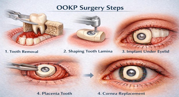
Figure 1: This image shows the steps of OOKP surgery.
Image Courtesy: Created by the Author
Recovery and Aftercare
Recovery requires patience. Patients spend time in the hospital after each stage and need months of antibiotic and steroid eye drops. (1) Lifelong follow-up with ophthalmologists and dental specialists is essential, and some patients may require glasses or contact lenses to optimise vision. (5)
Success Rates and Outcomes
Long-term studies report anatomical success rates of 85–94%, making OOKP one of the most successful options for carefully selected patients with end-stage corneal blindness. (1)
Many individuals regain functional vision, with some even achieving driving or reading vision. One remarkable example is Brent Chapman, who regained approximately 20/30 vision after being blind for two decades. (3)
Risks and Complications
OOKP is complex and carries significant risks at each stage.
During Surgery
It is one of the most complex eye surgeries ever performed. As with any major surgery, complications can occur, including infection, bleeding, and problems related to tooth extraction and healing. (1)
After Surgery
Long-term risks include graft failure, bone resorption, and glaucoma, which affects many patients and requires lifelong monitoring. Because the procedure restores only central vision, peripheral vision remains limited. Patients also need regular follow-up visits, lifelong lubricating eye drops, and ongoing specialist care. Owing to these challenges, OOKP is reserved only for patients with severe corneal blindness when all other treatment options have failed. (1,2)
Conclusion
OOKP (“tooth-in-eye”) surgery is an extraordinary procedure designed for the most severe corneal blind cases. By using a patient’s own tooth and bone to hold an artificial lens, it by passes the hostile eye surface. It works for people with no other options. While it entails multiple surgeries and carries risks (infection, glaucoma, graft failure).
References
- McManes (2024). Tooth in eye surgery. All about vision. https://www.allaboutvision.com/treatments-and-surgery/vision-surgery/corneal/tooth-in-eye-surgery/
- Upton J. (2025). A tooth for an eye : restoring sight when there is no hope. The ophthalmologist. https://theophthalmologist.com/issues/2025/articles/december/a-tooth-for-an-eye-restoring-sight-when-there-is-no-hope/
- Sheffield T. (2025). Blind man can now see after having his tooth implanted in his eye in science fictiony surgery. People. https://people.com/blind-man-can-now-see-after-rare-tooth-in-eye-surgery-
- Kaur J. (2018). Osteo-odonto keratoprosthesis: Innovative dental and ophthalmic blending. Journal of Indian Prosthodontic Society, 18(2), 89–95. https://doi.org/10.4103/jips.jips_283_17
- Koda A. (2025). Rare tooth-in-eye surgery restores man’s vision after two decades. Wral. https://www.wral.com/story/this-man-can-see-again-thanks-to-a-tooth-implanted-in-his-eye/22159859/
About the Author

Mohammad Akif Akram
Adjunct Optometrist,
Recent Comments